
Fascial Expansion: MSR Jaw Protocol
Updated: Sep 6

Fascial expansions are part of a continuous, three-dimensional connective-tissue network extending throughout the body. Far from being passive wrapping, fascia surrounds, separates, interweaves with, and connects muscles, bones, joints, nerves, blood vessels, and organs. Its collagen fibres and fluid-rich extracellular matrix adapt to movement and mechanical loading, while its sensory innervation contributes to proprioception, nociception, and motor coordination.
Research presented through the International Fascia Research Congress and the anatomical and histological work of Stecco and colleagues has expanded our understanding of fascia as a dynamic system. Myofascial expansions can transmit tension between adjoining structures, influence tissue gliding, and contribute to the coordination of movement across regions. These relationships provide a rationale for looking beyond the immediate site of jaw pain when clinically relevant findings suggest involvement of the cervical spine or other connected regions.
Traditional Chinese Medicine offers another way of interpreting relationships among different areas of the body. Its meridian system developed through centuries of clinical observation and should not be reduced to Western anatomical terminology. At the same time, research demonstrating relationships between many acupuncture points and connective-tissue planes raises intriguing questions about where these perspectives may intersect.
The MSR Jaw Protocol brings these bodies of knowledge together. Fascial assessment and manual treatment are combined with selected acupuncture or acupressure points, joint procedures, and functional exercise. The intention is not to impose one explanation on every case of TMD, but to identify meaningful patterns of tension, sensitivity, movement, and coordination that may be contributing to the individual’s symptoms.
Some of the proposed relationships can be demonstrated anatomically or measured physiologically. Others remain clinical models that continue to evolve as research methods improve. Used thoughtfully, these perspectives provide practitioners with a broader framework for addressing jaw pain, movement restrictions, and associated musculoskeletal dysfunction.
Article Index

Fascial Expansions and Traditional Chinese Medicine
Fascial expansions offer a compelling way to examine TMD within a broader system of anatomical continuity, force transmission, sensory input, and movement coordination. Traditional Chinese Medicine provides another perspective, developed through centuries of clinical observation, in which local and distant points are connected through channels that influence function across different regions of the body.
The MSR Jaw Protocol brings these perspectives together by integrating fascial anatomy, kinetic-chain relationships, and selected acupuncture or acupressure points, including ST6, ST7, SI8, LI4, and GB20. These points are combined with appropriate soft-tissue and osseous procedures, functional exercise, and patient education.
This approach does not assume that every person with TMD has the same pattern of dysfunction. The choice of local and distant treatment sites is guided by the history, physical examination, movement testing, palpation, and the principles of both fascial assessment and TCM. The objective is to address relevant structural and functional relationships rather than focusing exclusively on the location of pain.
Jaw-Related Fascial Planes
Fascia in the cranial, temporal, orbital, and pterygoid regions forms an intricate three-dimensional network. These tissues provide mechanical continuity, permit movement between adjacent structures, and contain sensory nerve endings that may contribute to proprioception and pain.
Manual therapy and acupuncture introduce mechanical and sensory input into this network through different methods. Their clinical effects may involve changes in tissue mobility, sensory processing, muscle activity, circulation, and pain modulation. Although research continues to investigate these mechanisms, the following fascial structures provide an important anatomical foundation for the MSR Jaw Protocol.
Epicranial fascia: The epicranial aponeurosis connects the frontalis and occipitalis muscles across the scalp and has relationships with the temporal fascia surrounding the temporalis muscle. Through its attachments and continuities, this system links movement and tension across the scalp, temple, and craniofacial region.
Tenon’s capsule and the orbital fascia: Anteriorly, the craniofacial fascial system continues into the complex fascial structures of the orbit. Tenon’s capsule surrounds the globe from near the optic nerve to the front of the eye, separating it from the surrounding orbital fat. It is pierced and reflected around the tendons of the extraocular muscles and blends posteriorly with tissues surrounding the optic nerve. These relationships demonstrate the close anatomical integration of the orbit with the surrounding craniofacial fascial system.
Pterygoid fascia: Fascial tissues surround and separate the medial and lateral pterygoid muscles within the infratemporal region. These tissues have relationships with the mandible, muscles of mastication, and connective tissues surrounding the TMJ. The lateral pterygoid plays an important role in mandibular translation and the coordination of the condyle-disc complex.
Attachments of the lateral pterygoid vary. Fibres may insert into the mandibular condyle, TMJ capsule, articular disc, or a combination of these structures. This variability means the muscle should not be described as controlling disc position in exactly the same way in every person. Nevertheless, its close relationship with the disc-capsule complex makes the lateral pterygoid and its surrounding fascia clinically relevant when assessing jaw movement, pain, clicking, or altered coordination.
These structures should not be viewed as isolated layers. They are parts of an integrated fascial environment that interacts continuously with muscles, joints, nerves, blood vessels, and sensory systems. This anatomical perspective helps explain why an MSR assessment may extend beyond the painful area and include the temporal region, cervical spine, shoulder girdle, upper limb, and selected distal points.

Acupuncture & Acupressure
for Jaw Pain
Traditional Chinese Medicine describes acupuncture points, or acupoints, as specific locations situated along channels that connect different regions and functions of the body. Within this framework, health depends partly on the balanced movement of Qi through these channels. Pain and impaired function may arise when this movement becomes disrupted, deficient, or stagnant.
Acupuncture uses fine needles to stimulate selected points, while acupressure applies manual pressure and tissue movement. Although the two approaches share point locations and traditional principles, they are not identical interventions and should not be assumed to produce precisely the same physiological effects.
From a TCM perspective, point selection is based on more than proximity to the painful area. Local points such as ST6 and ST7 may be combined with distal points such as LI4 or SI8 according to the channels involved, the pattern of symptoms, and findings elsewhere in the body. Treatment is intended to regulate the movement of Qi, reduce stagnation, restore balance, and support the body’s capacity for healing.
Contemporary research offers several possible physiological explanations for some of these effects. Many acupuncture points are located near nerves, motor points, blood vessels, connective-tissue planes, or interfaces between different tissue layers. Research has also demonstrated that needle manipulation can deform connective tissue and produce cellular responses in fibroblasts. Acupuncture may influence peripheral sensory input, spinal cord processing, central pain-modulating systems, endogenous opioids, local circulation, and autonomic regulation.
These findings do not establish that meridians are simply nerves, blood vessels, or fascial planes. TCM and contemporary biomedicine describe the body through different conceptual frameworks. Their points of convergence are scientifically intriguing, but neither framework needs to be reduced entirely to the other.
Acupuncture/Pressure Techniques
Acupuncture involves more than inserting a needle and leaving it motionless. Depending on the treatment approach, the practitioner may rotate, lift, thrust, or otherwise manipulate the needle to obtain a characteristic tissue response. The patient may experience heaviness, warmth, pressure, tingling, or a spreading sensation traditionally known as de qi. The practitioner may also perceive increased resistance around the needle, commonly described as needle grasp. These related responses are important within many acupuncture traditions, although they are not required in every technique or for every therapeutic effect.
Acupressure applies a different form of mechanical input. In the MSR protocol, the practitioner identifies the point, considers the underlying anatomy, and palpates the surrounding tissues for changes in sensitivity, density, mobility, or tension. Treatment may include sustained pressure, clockwise and counterclockwise movements, fascial kneading, tissue rolling, or multidirectional loading.
The selected point is not treated as an isolated spot. Its relationship with surrounding fascia, muscles, joints, nerves, and the broader channel or kinetic pattern is considered. Local points may therefore be combined with distal points when the assessment and TCM pattern suggest that these relationships are clinically relevant.
Possible effects include altered sensory input, pain modulation, changes in muscle activity, local circulatory responses, and mechanical signalling within connective tissue. Broader neural, autonomic, and immune effects continue to be investigated. Tensegrity offers an additional model for considering how tension and compression are distributed through an interconnected biological system, although it should be understood as a conceptual framework rather than a complete explanation of treatment effects.
By bringing together precise anatomy, skilled palpation, fascial continuity, and TCM channel theory, the MSR approach provides more than one lens through which to interpret and treat jaw pain. The purpose is not to force these perspectives into a single explanation, but to use each thoughtfully when it adds meaningful information to the individual assessment.
Specific Acupuncture Points
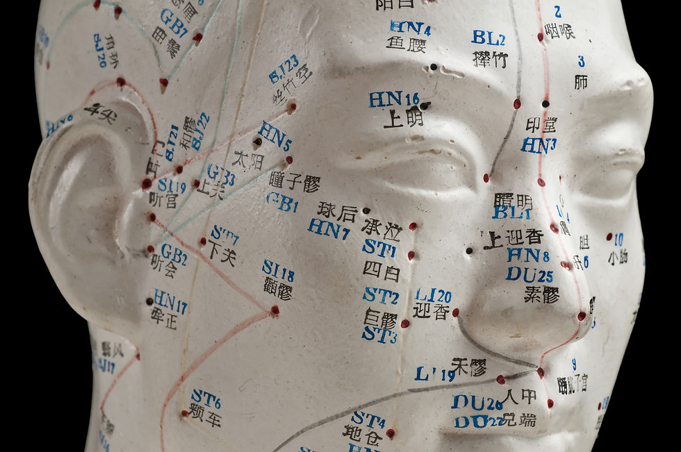
The MSR Jaw Protocol may incorporate ST6, ST7, SI8, LI4, and GB20. In Traditional Chinese Medicine, these points are selected according to channel relationships, the individual pattern of symptoms, and findings elsewhere in the body. Some are located close to the jaw, while others are distal points chosen for their broader relationships with the face, head, neck, shoulder, and upper limb.
From a contemporary perspective, stimulation of these points provides mechanical and sensory input to the skin, fascia, muscles, nerves, and other underlying tissues. Acupuncture research suggests that treatment may influence pain modulation, muscle activity, local circulation, connective-tissue signalling, and central nervous system processing. The physiological response to acupressure may overlap with some of these mechanisms, but acupuncture and acupressure should not be considered identical interventions.
The descriptions below present both the traditional rationale and the anatomical or musculoskeletal relevance of each point.

ST 6 (Jiache):
Location: ST6 is located over the prominence of the masseter muscle, approximately one finger-width anterior and superior to the angle of the mandible. Contracting the masseter can make the point easier to identify.
TCM perspective: ST6 is traditionally used to regulate Qi in the face and jaw, relieve pain, reduce swelling, and address difficulty opening the mouth, toothache, and facial dysfunction.
Anatomical and clinical relevance: ST6 provides direct access to the masseter region, which is frequently tender or overactive in people who clench or experience myogenous TMD. Treatment may be directed toward local sensitivity, muscle tone, fascial mobility, and sensory input.

ST 7 (Xiaguan):
Location: ST7 is situated anterior to the ear, in the depression below the zygomatic arch and anterior to the mandibular condyle when the mouth is closed.
TCM perspective: ST7 is traditionally used for jaw pain, restricted opening, toothache, tinnitus, facial pain, and facial paralysis. It is considered an important local point for disorders affecting the jaw and ear regions.
Anatomical and clinical relevance: ST7 lies close to the TMJ, masseter, lateral pterygoid region, branches of the trigeminal nerve, and adjacent fascial structures. Depending on how it is stimulated, it may provide input to tissues involved in jaw movement, pain, and coordination.

SI 8 (Xiaohai):
Location: SI8 is located on the medial side of the elbow, in the depression between the olecranon of the ulna and the medial epicondyle of the humerus, most easily identified with the elbow flexed.
TCM perspective: SI8 is the He-Sea point of the Small Intestine channel. It is traditionally used to clear heat, reduce swelling, ease pain, and address conditions involving the elbow, upper limb, shoulder, scapular region, neck, and channel pathway toward the cheek and ear.
Although SI8 is distant from the jaw, distal point selection is fundamental to TCM. Its inclusion reflects the course of the Small Intestine channel and the clinical pattern being treated rather than simple proximity to the painful area.
Anatomical and clinical relevance: Within the MSR protocol, SI8 may be considered when examination identifies meaningful relationships among the medial elbow, upper limb, shoulder girdle, cervical region, and jaw. This represents the integration of TCM channel theory with fascial and musculoskeletal assessment.
The ulnar nerve passes close to SI8. Needling or sustained manual pressure requires precise localization and appropriate training.

LI 4 (Hegu):
Location: LI4 is located on the back of the hand between the first and second metacarpal bones, near the midpoint of the second metacarpal. The first dorsal interosseous muscle forms much of the underlying tissue.
TCM perspective: LI4 is the Yuan-Source point of the Large Intestine channel and one of the most frequently used distal points for conditions affecting the face and mouth. It is traditionally selected to regulate Qi, relieve pain, clear heat, and address headache, facial pain, toothache, jaw discomfort, and stress-related tension.
Anatomical and clinical relevance: LI4 contains sensory input from branches of the radial and median nerve territories and may contribute to pain modulation through peripheral and central neural mechanisms. It is commonly combined with local facial points rather than used as an isolated treatment.
Traditional acupuncture teaching advises avoiding or using particular caution with strong stimulation of LI4 during pregnancy. Clinical decisions should follow current professional guidance and the practitioner’s regulated scope of practice.

GB 20 (Fengchi):
Location: GB20 is located below the occipital bone in the depression between the upper attachments of the sternocleidomastoid and trapezius muscles.
TCM perspective: GB20 is traditionally used to dispel wind, clear heat, benefit the head and eyes, and regulate the flow of Qi and blood. Its traditional indications include headache, dizziness, neck stiffness, visual symptoms, and certain ear-related complaints.
Anatomical and clinical relevance: GB20 lies within a complex suboccipital region containing muscles, fascia, sensory nerves, and important neurovascular structures. It may be considered when jaw symptoms coexist with cervical restriction, suboccipital sensitivity, or headache. Treatment may influence local sensory input, muscle tone, and cervical movement, which can be clinically relevant in some TMD presentations.
Deep needling in this region carries significant anatomical risk and should be performed only by practitioners with appropriate acupuncture training and a detailed understanding of the regional anatomy.
Safety and Point Selection
These points are examples within an integrated protocol, not a universal prescription for TMD. Point selection, depth, direction, intensity, and method of stimulation must be adapted to the individual and to whether acupuncture or acupressure is being used.
Needling should be avoided through local infections, open wounds, or compromised tissues. Additional precautions may be required for pregnancy, anticoagulant use, bleeding disorders, significant medical conditions, previous surgery, and anatomically sensitive regions.
Acupuncture needling should only be performed by practitioners who have the appropriate training and for whom acupuncture falls within their legally defined scope of practice. All procedures must comply with the laws, regulations, and professional standards of the practitioner’s jurisdiction.

Fascial Expansion Demonstration
In this video, Dr. Brian Abelson, DC, reviews the fascial planes and anatomical relationships involved in jaw function. He then demonstrates one way MSR integrates fascial assessment and manual treatment with selected acupuncture or acupressure points.
The demonstration illustrates how local and distal findings may be incorporated into an individualized treatment strategy. It represents one component of a broader approach that may also include joint procedures, exercise, patient education, and referral when appropriate.
Acupuncture needling should only be performed by practitioners with appropriate training and when it falls within their legally defined scope of practice.

Conclusion
Fascia is far more than passive wrapping. It forms a continuous, adaptable, and richly innervated system that contributes to force transmission, proprioception, pain, and movement coordination. In the craniofacial region, fascial relationships among the scalp, temple, orbit, muscles of mastication, TMJ, and cervical spine provide an important framework for understanding jaw function within a larger biological system.
Traditional Chinese Medicine offers another valuable perspective. Its use of local and distal points reflects centuries of clinical observation concerning relationships among different regions of the body. Contemporary research into connective-tissue planes, mechanotransduction, sensory modulation, and acupuncture does not fully explain the meridian system, but it reveals fascinating areas of convergence worthy of continued investigation.
The MSR Jaw Protocol integrates these perspectives through fascial assessment, manual treatment, acupuncture or acupressure, osseous procedures, and functional exercise. No single point or technique is appropriate for every person with TMD. The value lies in identifying clinically meaningful patterns and selecting interventions that fit the individual.
Fascial anatomy, TCM channel theory, and modern pain science offer different windows into the same extraordinarily complex human system. Used with precision, humility, and appropriate training, they can broaden how practitioners assess jaw pain and support the restoration of movement and function.
References
Antonopoulou, M., Iatrou, I., Paraschos, A., & Anagnostopoulou, S. (2013). Variations of the attachment of the superior head of the human lateral pterygoid muscle. Journal of Cranio-Maxillofacial Surgery, 41(6), e91–e97.
Deadman, P., Al-Khafaji, M., & Baker, K. (2007). A Manual of Acupuncture. Journal of Chinese Medicine Publications.
Di Francesco, F., Minervini, G., Siurkel, Y., Cicciù, M., & Lanza, A. (2024). Efficacy of acupuncture and laser acupuncture in temporomandibular disorders: A systematic review and meta-analysis of randomized controlled trials. BMC Oral Health, 24, 174.
Ha, S., Kang, S. W., & Lee, S. (2025). Comparative effectiveness of traditional East Asian medicine treatments for temporomandibular joint disorders: A systematic review and network meta-analysis. Integrative Medicine Research, 14(1), 101114.
Han, J. S. (2003). Acupuncture: Neuropeptide release produced by electrical stimulation of different frequencies. Trends in Neurosciences, 26(1), 17–22.
Kaptchuk, T. J. (2000). The Web That Has No Weaver: Understanding Chinese Medicine. Contemporary Books.
Krause, F., Wilke, J., Vogt, L., & Banzer, W. (2016). Intermuscular force transmission along myofascial chains: A systematic review. Journal of Anatomy, 228(6), 910–918.
Kumka, M., & Bonar, J. (2012). Fascia: A morphological description and classification system based on a literature review. Journal of the Canadian Chiropractic Association, 56(3), 179–191.
Langevin, H. M. (2021). Fascia mobility, proprioception, and myofascial pain. Life, 11(7), 668.
Langevin, H. M., Churchill, D. L., & Cipolla, M. J. (2001). Mechanical signaling through connective tissue: A mechanism for the therapeutic effect of acupuncture. FASEB Journal, 15(12), 2275–2282.
Langevin, H. M., Bouffard, N. A., Badger, G. J., Churchill, D. L., & Howe, A. K. (2006). Subcutaneous tissue fibroblast cytoskeletal remodeling induced by acupuncture: Evidence for a mechanotransduction-based mechanism. Journal of Cellular Physiology, 207(3), 767–774.
Langevin, H. M., & Yandow, J. A. (2002). Relationship of acupuncture points and meridians to connective-tissue planes. The Anatomical Record, 269(6), 257–265.
Lim, R., & Morrison, J. G. (1998). Tenon’s capsule: A scanning electron microscope study of the fascial planes around the rectus muscles. Australian and New Zealand Journal of Ophthalmology, 26(1), 29–35.
Meyer, D. R., Linberg, J. V., Wobig, J. L., & McCormick, S. A. (1991). Anatomy of the orbital septum and associated eyelid connective tissues. Ophthalmic Plastic and Reconstructive Surgery, 7(2), 104–113.
Park, E. Y., Cho, J. H., Lee, S. H., Kim, K. W., Ha, I. H., & Lee, Y. J. (2023). Is acupuncture an effective treatment for temporomandibular disorder? A systematic review and meta-analysis of randomized controlled trials. Medicine, 102(38), e34950.
Pratt, R. L. (2021). Hyaluronan and the fascial frontier. International Journal of Molecular Sciences, 22(13), 6845.
Schleip, R., Adstrum, S., Hedley, G., Stecco, C., & Yucesoy, C. A. (2019). Fascial nomenclature: Update on the related consensus process. Clinical Anatomy, 32(7), 929–933.
Schmolke, C. (1994). The relationship between the temporomandibular joint capsule, articular disc, and jaw muscles. Journal of Anatomy, 184(2), 335–345.
Stecco, C. (2015). Functional Atlas of the Human Fascial System. Elsevier.
Stecco, C., & Schleip, R. (2016). A fascia and the fascial system. Journal of Bodywork and Movement Therapies, 20(1), 139–140.
Stecco, C., Stern, R., Porzionato, A., Macchi, V., Masiero, S., Stecco, A., & De Caro, R. (2011). Hyaluronan within fascia in the etiology of myofascial pain. Surgical and Radiologic Anatomy, 33(10), 891–896.
Suárez-Rodríguez, V., Fede, C., Pirri, C., Petrelli, L., Loro-Ferrer, J. F., Rodríguez-Ruiz, D., De Caro, R., & Stecco, C. (2022). Fascial innervation: A systematic review of the literature. International Journal of Molecular Sciences, 23(10), 5674.
Vickers, A. J., Vertosick, E. A., Lewith, G., MacPherson, H., Foster, N. E., Sherman, K. J., et al. (2018). Acupuncture for chronic pain: Update of an individual patient data meta-analysis. The Journal of Pain, 19(5), 455–474.
Article Index
Disclaimer:
The articles and embedded videos on the MSR website are provided for educational and informational purposes only. They are not a substitute for individualized assessment, diagnosis, or treatment by an appropriately qualified healthcare professional.
Some videos demonstrate clinical examination and manual therapy procedures intended for licensed or regulated healthcare professionals with appropriate training. These procedures should be performed only within the practitioner’s legal scope of practice and should not be attempted as self-treatment.
Viewing or using this content does not establish a practitioner-patient relationship. If you have questions about a medical condition or symptoms that are new, severe, or worsening, consult an appropriately qualified healthcare provider.
MSR is not responsible for decisions made or actions taken solely on the basis of this educational content. For complete information about website use, third-party links, privacy, and legal terms, please review the MSR Terms of Use, Privacy Policy, and full website disclaimer.
Article Index
DR. BRIAN ABELSON, DC. - The Author

Dr. Brian Abelson is a chiropractor, author, and educator with more than 30 years of clinical experience in musculoskeletal care, rehabilitation, and sports performance. His educational work focuses on musculoskeletal conditions, human movement, sports biomechanics, and evidence-informed approaches to treatment and rehabilitation.
He is the developer of Motion Specific Release (MSR) and Clinical Director of Kinetic Health in Calgary, Alberta, Canada.
Article Index

Continue Learning with MSR Courses and MSR Pro
For Healthcare Practitioners
Motion Specific Release integrates assessment, clinical reasoning, manual therapy, exercise, and patient education within one adaptable approach to musculoskeletal care.
MSR Pro and MSR courses support different parts of that learning process:
Structured Clinical Education: MSR courses connect applied anatomy and biomechanics with orthopedic and neurological assessment, myofascial procedures, fascial expansions, joint mobilization, manipulation, and rehabilitation.
Hands-On Skill Development: Live training provides opportunities to refine palpation, patient positioning, force application, and clinical decision-making through practice and direct feedback.
MSR Pro Clinical Library: Members receive access to more than 200 MSR procedures, downloadable and fillable clinical forms, examination resources, rehabilitation progressions, and exercise-prescription materials.
Extensive Video Resources: More than 800 videos support examination, treatment procedures, exercise rehabilitation, and clinical application.
Patient Education: A library of 50 condition articles helps practitioners explain common musculoskeletal problems and provide patients with practical information.
Continued Development: Resources are updated as MSR courses and clinical education continue to evolve.
Videos and articles can introduce concepts, but hands-on proficiency develops through deliberate practice, feedback, and experience. MSR is designed to help practitioners connect technique with clinical reasoning and adapt care to the individual rather than apply the same protocol to everyone.
Explore MSR Pro | View Online and Live Courses


Comments