The Fascinating World of Fascia: Enhancing Treatments with Customized Techniques
- Dr. Brian Abelson DC
- Apr 18, 2023
- 11 min read
Updated: Feb 25, 2024

Incorporating "specific customized" fascial release techniques into your existing practices can lead to remarkable outcomes(1).To appreciate the significance of fascia, let's first examine its multiple functions. Fascia is crucial for communication, preserving our body's history, and serving as both a tensional network and a living matrix.
Article Index
What Makes Fascia So Unique
Communication
While it is common knowledge that the nervous system acts as our body's communication network, recent research has revealed that the fascial system contains ten times the number of sensory nerve receptors compared to those innervating muscles(1)(2)(3). These receptors include various types, such as myelinated proprioceptive endings (Golgi, Paccini, and Ruffini) and unmyelinated free nerve endings. This information has shifted our understanding of fascia from a static wrapping substance to the realization that, given the astonishing number of nerve receptors, fascia is the body's most crucial perceptual organ(1)(2)(3).

Fascia Holds Our History
Our fascial network serves as a record of our life experiences! Each injury or physical force we encounter sends mechanical forces throughout our body. Over time, these forces lead to transcriptional (RNA) changes, which in turn alter our fascial architecture(4). Such changes can result in imbalances, adhesion formation, thickening, or decreased mobility. It's fascinating to consider how mechanical forces initiate transcription – the process of creating an RNA copy of a gene sequence and producing corresponding proteins – based on our body's physical history. As practitioners, we can gather a wealth of information from fascia, which helps us develop suitable and effective treatments for our patients.
The Tensional Network
Fascia is often described as "one interconnected tensional network that adapts its fiber arrangement and density according to local tensional demands"(5). When fascial tension is well-balanced, it distributes force throughout the body and allows us to store and release energy for propulsion. However, when fascial tension is imbalanced, hypertensive, or restricted, it can become the source of various dysfunctions.
The image above, copyrighted by Professor Jaap Van Der Wal, MD, PhD University Maastricht, whom I met at the Second International Fascia Congress in 2009, displays the proximal lateral elbow region with the muscles dissected away. Considering the convergence of all surrounding fascial fibers into the lateral elbow and their interrelation, it's easy to understand the concept of a tensional network. Extending this view to encompass the entire body reveals the crucial role fascia plays in all movement.
Fascia as a Living Matrix
Fascia is not a simple packaging material enclosing our internal organs. Rather, it is a living matrix that surrounds, supports, and penetrates every muscle, tendon, ligament, bone, joint, cardiovascular, and neurological structure in the body.
Fascia is a dynamic web that maintains tension for force transmission, shock absorption, and communication. In essence, your fascial network represents the ultimate physical manifestation of a kinetic chain. These functions are closely linked to the cells contained within the fascia(6).
Though individual cells make up only a small portion of the total fascial material, some cells play crucial roles in architectural design, repair, and fascial tension regulation. These dynamic cells are known as fibroblasts.

Fibroblasts are crucial because they form the foundation of the fascial system. These dynamic cells can change in length within moments when fascial tissue lengthens under compressive loads. What's remarkable about fibroblasts is not only their ability to change shape, but also their capacity to transform into another cell type called myofibroblasts. Myofibroblasts can contract and directly affect fascial tension, which in turn influences force transmission, energy storage, and communication. The contractibility of myofibroblasts is four times stronger than that of regular fibroblasts(7) (Fibroblast images: https://www.youtube.com/watch?v=kgnp0ZU51vc).
Myofibroblasts form when mechanical strain increases in the body, which may result from injury, repetitive stress, muscle imbalances, or even insufficient physical activity. Myofibroblasts play both positive and negative roles. For instance, they are vital in wound healing, but they also contribute to fascial contractures and scar tissue formation in conditions such as frozen shoulder or Dupuytren's contracture(7).

The connection between increased myofascial tension, chronic anxiety (heightened sympathetic nervous system activity), and myofibroblasts has long intrigued researchers. Although initially unclear, studies have now established a link between myofibroblasts, the sympathetic nervous system, and small proteins called cytokines(8).
Initially, researchers hypothesized that increased myofibroblast contraction was associated with sympathetic neurotransmitters (epinephrine, adrenaline, and acetylcholine), but this was later disproven. Instead, they discovered that the cytokine TGF-B1, produced during heightened sympathetic activity (stress), was the connecting factor. TGF-B1 results in significant myofibroblast contraction, leading to increased myofascial tension and related dysfunctions(7)(9).
From a practitioner's perspective, this is crucial information. Balancing the fascial system could positively affect myofibroblast activity, potentially reducing overall myofascial tension. For instance, performing procedures such as the MSR™ Diaphragmatic Release, combined with appropriate breathing exercises, may quickly decrease overall sympathetic nervous system activity. Integrating this approach into treatments could offer significant relief for numerous patients experiencing chronic pain and various stress-related conditions.
Fascial Expansions

Fascial expansions refer to the complex network of interconnected fascial planes that span various anatomical structures, such as the jaw, shoulder, hip, and knee. Fascia, a specialized connective tissue composed primarily of collagen, envelops and interpenetrates muscles, bones, nerves, blood vessels, and organs, providing structural support and facilitating biomechanical communication between these components [7].
Fascial planes are dynamic, adaptive structures that respond to the mechanical forces exerted on them, altering their density and fiber arrangement accordingly. This interconnectedness allows for the efficient transmission and distribution of forces throughout the body, which is crucial for maintaining proper biomechanics and musculoskeletal health [6]. Moreover, the fascial system's intricate connections contribute to the body's ability to store and release energy during movement and athletic performance [23].
Understanding the interconnected nature of fascial planes provides valuable insights into the causes and treatment of numerous musculoskeletal conditions. For instance, restrictions or imbalances in one fascial plane may result in compensatory changes and dysfunctions in other interconnected regions, leading to pain and movement limitations. Consequently, therapeutic approaches that target the fascial system, such as manual therapies, myofascial release techniques, and acupuncture, can be particularly effective in addressing musculoskeletal issues by restoring balance and promoting optimal force transmission throughout the body [24].
Fascial Expansions and Traditional Chinese Medicine (TCM)
For example, addressing temporomandibular joint disorders (TMJ/TMD) by leveraging fascial expansions provides a well-rounded method that brings together the latest fascial research[1], the understanding of kinetic chain connections[2], and the time-tested principles of acupuncture or traditional Chinese medicine[3]. In this article, we'll explore how fascial layers interact with acupuncture points ST6, ST7, ST8, SI8, LI4, and GB20[4][5]. To achieve the best outcomes, it's important to combine this approach with both soft tissue and osseous techniques[6], as well as incorporating a routine of functional exercises[7].
Jaw-Related Fascial Planes
In the case of jaw pain, addressing restrictions in fascial planes is possible through various techniques, such as acupuncture and hands-on manipulation (soft tissue and skeletal procedures)[3][6]. The fascial planes outlined below play a significant role in jaw function, making it essential to consider these primary regions:
Epicranial Fascia: This fascinating fascia connects the occipitalis and frontalis muscles, seamlessly extending to the temporal fascia that wraps around the temporalis muscle[10]. As you move forward, the epicranial fascia transforms into Tenon's fascia[11].
Tenon's Fascia: Acting as a protective sheath, Tenon's fascia surrounds the levator muscle of the upper eyelid[11]. Interestingly, the rear third of Tenon's fascia combines with the orbital fat, ultimately connecting to the optic nerve's protective covering[12].
Pterygoid Fascia: Encompassing the medial and lateral pterygoid muscles, the pterygoid fascia attaches to the temporomandibular joint (TMJ) capsule[13]. A part of the upper head of the lateral pterygoid muscle directly inserts into the anteromedial region of the articular disc[14]. Consequently, the lateral pterygoid muscle and its associated fascia can have a direct effect on the articular disc's position during TMJ movement[13].
Image: Stecco, Carla; Stecco, Carla. Functional Atlas of the Human Fascial System (p. 109). Elsevier Health Sciences. Kindle Edition. I highly recommend this atlas, just click the link or the above image.
Acupressure

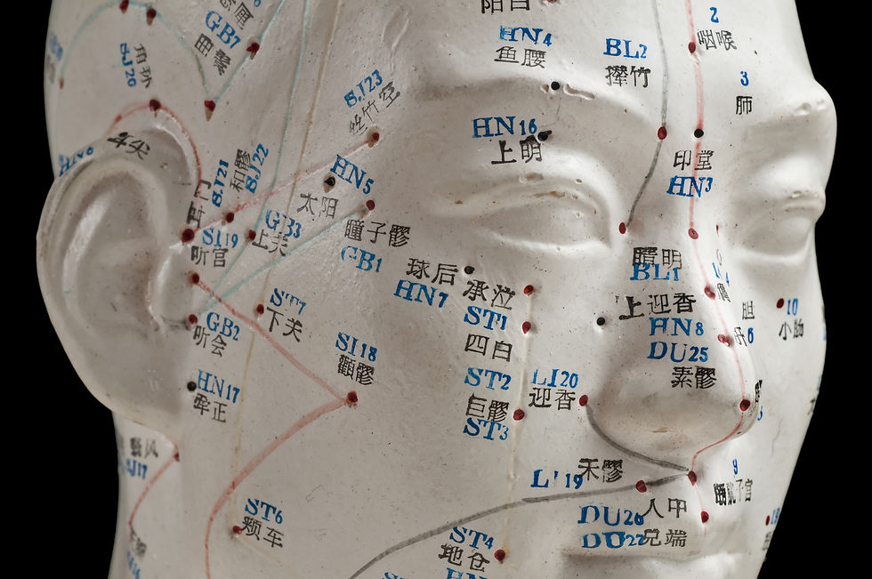
Acupuncture points, also known as acupoints or simply points, are specific locations on the body identified in Traditional Chinese Medicine (TCM) to possess therapeutic effects when stimulated[16]. These points are located along meridians or channels, believed to be pathways of energy flow called "Qi" (pronounced "chi") throughout the body[15]. According to TCM, stimulating acupuncture points can help restore balance, regulate the flow of Qi, alleviate pain, and promote healing in the body[16].
Recent research has shown that acupuncture points often correspond to areas with a high density of nerve endings, blood vessels, and lymphatic vessels, as well as increased electrical conductivity[17]. This suggests that the stimulation of acupuncture points may have physiological effects, such as the release of endorphins, neurotransmitters, and other pain-relieving substances[18], as well as the regulation of blood flow and the immune system[6].
When it comes to acupuncture techniques, needles are not merely inserted; they are rotated and pulled back and forth until the acupuncturist perceives a response in the tissue (sometimes referred to as a tug response)[3].
Acupressure
Acupressure follows a similar approach: stimulating a region to activate the nervous system and release tension within a fascial network of interconnected tissue[20].

Specific Acupuncture Points
We will continue with the example of jaw pain. In Traditional Chinese Medicine (TCM), acupuncture points ST6, ST7, ST8, SI8, LI4, and GB20 are commonly used to alleviate pain associated with temporomandibular joint disorders[21].
Note: We employ the same point when utilizing acupressure instead of acupuncture. The choice of technique depends on your professional scope of practice. It's crucial to remember that we're not only working on the acupuncture point but also releasing the surrounding fascia.
The location of these points is often described using the Chinese term "cun," a unit of measurement employed in acupuncture for locating points on the body[4]. One cun is approximately equal to the width of the patient's thumb at the knuckle[4]:
ST 6 (Jiache):
Location: At the prominence of the masseter muscle, one finger-width anterior and superior to the angle of the mandible.
Indications: Facial paralysis, trigeminal neuralgia, toothache, and temporomandibular joint disorders.
ST 7 (Xiaguan):
Location: Anterior to the ear, in the depression between the zygomatic arch and the mandibular notch.
Indications: Facial paralysis, temporomandibular joint disorders, toothache, and tinnitus.
SI 8 (Xiaohai):
Location: On the medial aspect of the elbow, in the depression between the olecranon process of the ulna and the medial epicondyle of the humerus.
Indications: Elbow pain, upper limb disorders, and conditions affecting the scapular and shoulder regions.
LI 4 (Hegu):
Location: Dorsal aspect of the hand, between the first and second metacarpal bones, approximately at the midpoint of the second metacarpal bone.
Indications: Headaches, toothaches, facial pain, neck pain, and various conditions related to the face and head.
GB 20 (Fengchi):
Location: On the posterior aspect of the neck, below the occipital bone, in the depression between the upper portion of the sternocleidomastoid and trapezius muscles.
Indications: Headaches, migraines, neck pain, dizziness, and conditions affecting the eyes and ears.
Fascial Expansion Demonstration
In this video, Dr. Abelson discusses the fascial planes that directly impact jaw function. He then demonstrates how practitioners can combine this knowledge with Traditional Chinese Medicine (Acupuncture/Acupressure). By understanding the interconnected nature of fascial planes and their effect on jaw function, along with the specific acupuncture points and techniques used in TCM, practitioners can effectively alleviate pain and foster healing for patients dealing with TMJ/TMD.
Conclusion
The fascial network, abundant with sensory nerve receptors, is pivotal for communication and biomechanical functions, evolving beyond its once-perceived role as a mere passive structure. Its adaptability to mechanical forces, capability to store and release kinetic energy, and its intricate interplay with acupuncture points underscore its significance in holistic health practices. By fusing traditional acupuncture methods with contemporary fascial insights, practitioners can enhance therapeutic outcomes.
The nuanced relationships between fascial planes, dynamic cells like fibroblasts and myofibroblasts, and their impact on fascial tension emphasize the integrated nature of our physiological systems. For health professionals, this interconnectedness highlights the necessity of a comprehensive approach, considering the entire tensional network. Through a deep understanding and utilization of these connections, we can promote optimal health, functionality, and well-being for patients.
DR. BRIAN ABELSON DC. - The Author

Dr. Abelson's approach in musculoskeletal health care reflects a deep commitment to evidence-based practices and continuous learning. In his work at Kinetic Health in Calgary, Alberta, he focuses on integrating the latest research with a compassionate understanding of each patient's unique needs. As the developer of the Motion Specific Release (MSR) Treatment Systems, he views his role as both a practitioner and an educator, dedicated to sharing knowledge and techniques that can benefit the wider healthcare community. His ongoing efforts in teaching and practice aim to contribute positively to the field of musculoskeletal health, with a constant emphasis on patient-centered care and the collective advancement of treatment methods.

Revolutionize Your Practice with Motion Specific Release (MSR)!
MSR, a cutting-edge treatment system, uniquely fuses varied therapeutic perspectives to resolve musculoskeletal conditions effectively.
Attend our courses to equip yourself with innovative soft-tissue and osseous techniques that seamlessly integrate into your clinical practice and empower your patients by relieving their pain and restoring function. Our curriculum marries medical science with creative therapeutic approaches and provides a comprehensive understanding of musculoskeletal diagnosis and treatment methods.
Our system offers a blend of orthopedic and neurological assessments, myofascial interventions, osseous manipulations, acupressure techniques, kinetic chain explorations, and functional exercise plans.
With MSR, your practice will flourish, achieve remarkable clinical outcomes, and see patient referrals skyrocket. Step into the future of treatment with MSR courses and membership!
References
Mitchell JH, and Schmidt RF. (1977). Cardiovascular reflex control by afferent fibers from skeletal muscle receptors. In: Shepherd JT, et al, eds, Handbook of physiology, Section 2, Vol. III, Part 2. Bethesda: American Physiological Society, pp. 623-658.
Schleip R. (2003). Fascial plasticity— a new neurobiological explanation. Part 1. J Bodyw Mov Ther, 7(1), pp. 11-19.
Van der Wal J. (2009). The architecture of the connective tissue in the musculoskeletal system: An often-overlooked functional parameter as to proprioception in the locomotor apparatus. In: Huijing PA, et al, eds. Fascia research II: Basic science and implications for conventional and complementary health care. Munich: Elsevier GmbH.
Chen C, and Ingber D. (2007). Tensegrity and mechanoregulation: from skeleton to cytoskeleton. In: Findley T, and Schleip R, eds. Fascia research. Oxford: Elsevier, pp. 20-32.
Findley T, and Schleip R. (2009). Introduction. In: Huijing PA, Hollander P, Findley TW, and Schleip R, eds. Fascia research II. Basic science and implications for conventional and complementary health care. München: Urban and Fischer.
Schleip R, Findley TW, Leon Chaitow L, and Huijing PA. (2012). Fascia: The Tensional Network of the Human Body - E-Book: The science and clinical applications in manual and movement therapy. Canada: Elsevier
Schleip R, Klingler W, and Lehmann-Horn F. (2006). Fascia is able to contract in a smooth muscle-like manner and thereby influence musculoskeletal mechanics. In: Liepsch D, ed. 5th World Congress of Biomechanics, Munich (Germany) 29 July– August 4, 2006. Bologna: Medimond International Proceedings, pp. 51-54.
Bhowmick S, Singh A, Flavell RA, et al. (2009). The sympathetic nervous system modulates CD4(+) FoxP3(+) regulatory T cells via a TGF-beta-dependent mechanism. J Leukoc Biol, 86, pp. 1275-1283.
Stefano GB, and Esch T. (2005). Integrative medical therapy: examination of meditation’s therapeutic and global medicinal outcomes via nitric oxide (review). Int J Mol Med, 16(4), pp. 621-630.
Yahia LH, Pigeon P, and DesRosiers EA. (1993). Viscoelastic properties of the human lumbodorsal fascia. J Biomed Eng, 15(5), pp. 425-429.
Langevin HM. Fibroblast cytoskeletal remodeling contributes to viscoelastic response of arealoar connective tissue under uniaxial tension. [DVD Recording] Boston MA: Second International Fascia Research Congress; 2009.
Sahara W, Sugamoto K, Murai M, et al: Three-dimensional clavicular and acromioclavicular rotations during arm abduction using vertically open MRI. J Orthop Res 25:1243, 2007.
Article Index
Disclaimer:
The content on the MSR website, including articles and embedded videos, serves educational and informational purposes only. It is not a substitute for professional medical advice; only certified MSR practitioners should practice these techniques. By accessing this content, you assume full responsibility for your use of the information, acknowledging that the authors and contributors are not liable for any damages or claims that may arise.
This website does not establish a physician-patient relationship. If you have a medical concern, consult an appropriately licensed healthcare provider. Users under the age of 18 are not permitted to use the site. The MSR website may also feature links to third-party sites; however, we bear no responsibility for the content or practices of these external websites.
By using the MSR website, you agree to indemnify and hold the authors and contributors harmless from any claims, including legal fees, arising from your use of the site or violating these terms. This disclaimer constitutes part of the understanding between you and the website's authors regarding the use of the MSR website. For more information, read the full disclaimer and policies in this website.


Comments