
Knee Ligament Injuries: ACL, PCL, MCL and LCL
Updated: Sep 29

Knee ligament injuries can range from a mild sprain to a complete tear and may affect athletes as well as people injured during work, recreation, falls, or everyday activities. Depending on the ligament involved and the severity of the injury, symptoms can include pain, swelling, reduced movement, instability, or difficulty trusting the knee during activity.
A careful diagnosis is an important first step. This usually begins with the history of the injury and a thorough clinical examination. X-rays or MRI may be recommended when clinically indicated, particularly when a more significant ligament injury or associated damage to structures such as the meniscus or cartilage is suspected.
Treatment depends on which ligament is injured, the severity of the injury, associated injuries, the degree of knee instability, and the person's activity goals.
Many knee ligament injuries can be managed without surgery, using progressive rehabilitation to restore movement, strength, stability, and function. Manual therapy may also be used as an adjunct when appropriate. Other injuries, particularly some complete tears or complex multi-ligament injuries, may require orthopaedic consultation and surgical management.
The goal is not simply to treat an injured ligament. It is to restore a stable, capable knee and help you return safely to the activities that matter to you.
Article Index:
Anatomy of the Knee
The knee relies on four primary ligaments for passive stability: the anterior cruciate ligament (ACL), posterior cruciate ligament (PCL), medial collateral ligament (MCL), and lateral collateral ligament (LCL). Together with the joint surfaces, menisci, capsule, muscles, and neuromuscular system, these structures control anterior-posterior translation, varus-valgus motion, and rotation of the tibiofemoral joint.
Anterior Cruciate Ligament (ACL)
Origin: Anterior intercondylar area of the tibia.
Insertion: Posteromedial aspect of the lateral femoral condyle.
Primary Function: The ACL is the primary restraint to anterior translation of the tibia relative to the femur and contributes substantially to rotational stability. It also helps limit excessive hyperextension.
Common Injury Mechanisms: Rapid deceleration, cutting or pivoting, sudden changes of direction, and awkward landings are common non-contact mechanisms. Contact injuries may also occur.
Posterior Cruciate Ligament (PCL)
Origin: Posterior intercondylar area of the tibia.
Insertion: Lateral surface of the medial femoral condyle.
Primary Function: The PCL is the primary restraint to posterior translation of the tibia relative to the femur and also contributes to rotational stability.
Common Injury Mechanisms: A posteriorly directed force against the proximal tibia with the knee flexed is characteristic, such as a dashboard injury. Falls onto a flexed knee and sports-related hyperflexion or contact mechanisms may also injure the PCL.
Medial Collateral Ligament (MCL)
Origin: Medial femoral epicondylar region.
Insertion: The superficial MCL extends distally along the medial tibia, while deeper fibres have attachments closely associated with the joint capsule and medial meniscus.
Primary Function: The MCL complex is the primary restraint to excessive valgus stress and also contributes to rotational stability.
Common Injury Mechanisms: A force applied to the lateral knee producing valgus stress is common, although non-contact valgus and rotational mechanisms also occur.
Lateral Collateral Ligament (LCL)
Origin: Lateral femoral epicondyle.
Insertion: Fibular head.
Primary Function: The LCL is an important restraint to excessive varus stress and functions as part of the broader posterolateral corner (PLC) stabilizing complex.
Common Injury Mechanisms: Varus loading, hyperextension, and rotational trauma can injure the LCL. Clinically significant LCL injuries should raise consideration of associated posterolateral corner injury.
Passive Stability and Neuromuscular Control
The four major ligaments do not function as simple static restraints. Their tension changes throughout knee flexion and extension, and individual fibre bundles may contribute differently depending on joint position and loading.
Knee stability therefore results from the interaction between passive restraints and dynamic neuromuscular control. The cruciate and collateral ligaments constrain excessive translation and rotation, while the quadriceps, hamstrings, gastrocnemius, hip musculature, and other structures contribute active control during movement.
Ligaments also contain mechanoreceptors that contribute to proprioception and sensorimotor control. Following ligament injury, altered mechanical stability may therefore be accompanied by changes in proprioception, muscle activation, and movement strategies.
This interaction becomes particularly important during rehabilitation. Restoring function involves more than addressing the injured ligament itself. Strength, mobility, proprioception, balance, neuromuscular control, progressive loading, and task-specific movement may all need to be considered.
Examination of Knee Ligament Injuries
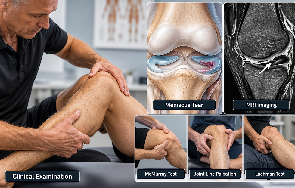
A thorough examination is essential when evaluating a suspected knee ligament injury. The objective is not simply to identify the painful structure, but to determine which ligament or combination of structures may be involved, the degree of instability, and whether associated meniscal, articular, neurological, or other injuries should be considered.
The examination begins with the history and mechanism of injury. Important findings may include an audible or perceived “pop,” rapid swelling, difficulty weight-bearing, episodes of giving way, locking or catching, and the patient's ability to continue activity immediately after the injury.
Physical examination typically progresses through observation, palpation, assessment of effusion and range of motion, neurovascular screening when indicated, and comparison of ligamentous stability with the opposite knee.
Specific ligament testing may include:
ACL: Lachman test, anterior drawer test, and pivot-shift testing.
PCL: Posterior drawer test, posterior sag sign, and other tests of posterior instability.
MCL: Valgus stress testing, commonly assessed at approximately 0° and 30° of knee flexion.
LCL and posterolateral structures: Varus stress testing at approximately 0° and 30°, with additional posterolateral testing such as the dial test when clinically indicated.
The examination should also consider meniscal injury, patellofemoral structures, joint-line tenderness, muscle function, and other potential sources of knee pain. Significant trauma or suspected multi-ligament injury requires particular attention to neurovascular status. Current ACL guidelines specifically include distal perfusion and tibial/peroneal nerve function in the examination.
Clinical examination may provide substantial diagnostic information, but imaging can be important when the diagnosis remains uncertain or associated injury is suspected. MRI can help confirm ligament injury and identify concomitant meniscal, cartilage, or other soft-tissue pathology.

Knee Examination - Effective Orthopaedic Testing
In this video, Dr. Brian Abelson demonstrates a systematic approach to knee examination. The assessment progresses from observation and palpation through range-of-motion testing and specific orthopaedic tests for ligamentous and meniscal injury.
The procedures include examination of the ACL, PCL, MCL, and LCL, along with tests used when evaluating other common causes of knee pain. The objective is to demonstrate how individual examination findings can be combined with the history and mechanism of injury to develop a more complete clinical picture.
Classification of Knee Ligament Injuries
Knee ligament injuries are commonly classified into three grades based on the extent of tissue disruption and resulting ligamentous laxity. Grading provides useful information about injury severity, but treatment decisions should also consider which ligament is involved, the degree of functional instability, associated injuries, activity demands, and the patient's goals.
Grade 1: Mild Sprain. The ligament remains structurally intact but has sustained mild fibre injury. There is typically localized tenderness with little or no measurable joint instability. Grade 1 injuries are generally managed conservatively with appropriate protection followed by progressive rehabilitation.
Grade 2: Partial Tear. A greater proportion of ligament fibres is disrupted, producing varying degrees of pain, swelling, and increased laxity while some structural continuity remains. Many Grade 2 injuries can be managed nonoperatively, although rehabilitation and protection strategies depend on the ligament involved and the degree of instability.
Grade 3: Complete Tear. The ligament is completely disrupted, often producing substantial laxity or instability. A Grade 3 injury does not automatically require surgery. Management depends on the ligament involved, whether the injury is isolated or part of a multi-ligament injury, the degree of functional instability, associated meniscal or cartilage injury, and the patient's occupational or athletic demands.
For example, isolated high-grade MCL injuries are often initially managed nonoperatively, while significant LCL/posterolateral corner injuries are more likely to require surgical consultation. Isolated PCL injuries, including selected high-grade injuries, may also respond well to structured nonoperative management, although high-grade instability, combined injuries, and the demands of the individual influence the decision.
Conservative Management of Ligament Injuries

Many knee ligament injuries can be managed successfully without surgery, but conservative care should be based on the ligament involved, severity of injury, degree of instability, associated injuries, and the functional demands of the patient.
The primary objectives are to protect the injured structures when necessary, restore knee motion, progressively rebuild strength and neuromuscular control, and gradually increase the knee's capacity to tolerate the demands of daily activity, work, or sport.
Rehabilitation forms the foundation of conservative management. Depending on the individual and stage of recovery, care may include activity modification, bracing or temporary use of crutches, progressive weight-bearing, range-of-motion exercise, strengthening, proprioceptive and neuromuscular training, and progressive return to functional activity.
Manual approaches, including Motion Specific Release (MSR), joint mobilization, massage, and other soft-tissue techniques, may be incorporated when clinically appropriate to address pain, movement limitations, and surrounding musculoskeletal findings. These interventions should complement, rather than replace, progressive rehabilitation.
Acute Stage of Ligamentous Injury Management
Immediately following a significant knee ligament injury, the priority is to identify the extent of injury, protect vulnerable structures, manage symptoms, and determine whether further investigation or referral is required.
Early management may include:
Protection: Temporarily reduce or modify activities that significantly increase pain or instability. Depending on the injury, bracing or crutches may be appropriate. Complete or prolonged rest is generally avoided when safe movement and loading can be introduced.
Progressive Weight-Bearing: Weight-bearing and movement can often be introduced progressively as symptoms and ligament stability permit. The appropriate rate depends heavily on the ligament injured and whether other structures are involved.
Compression: A compression sleeve or elastic bandage may help manage swelling and provide short-term comfort, although evidence for compression as an isolated treatment is limited.
Elevation: Elevation may be used as a simple strategy for managing swelling, although the evidence supporting a direct effect on tissue healing is limited. \
Cold Therapy: Ice can provide short-term analgesia for some patients, but it should not be presented as necessary for ligament healing. Evidence supporting cryotherapy as a stand-alone treatment for acute soft-tissue injuries is limited.
Early Movement: When clinically appropriate, restoring knee extension and flexion and beginning controlled muscle activation can help limit unnecessary stiffness and deconditioning.
Progressive Loading: As the acute symptoms settle, progressively increasing mechanical load becomes an important part of rehabilitation. Appropriate loading contributes to tissue adaptation and restoration of neuromusculoskeletal capacity.
The Ligament Matters
There is no single conservative protocol that applies equally to an ACL, PCL, MCL, or LCL injury.
For example, rehabilitation after an MCL injury may allow relatively early progressive loading while protecting the knee from excessive valgus stress. PCL rehabilitation requires consideration of posterior tibial translation and loading strategies that may stress the healing PCL. ACL rehabilitation places substantial emphasis on restoring range of motion, quadriceps strength, neuromuscular control, and functional stability.
Significant LCL or posterolateral corner injuries require particular caution because they may represent more complex instability patterns.
Conservative management is therefore not simply a decision to “avoid surgery.” It is an active, progressive rehabilitation process based on the injured structure, the patient's response, and the functional demands they need to regain.
Manual Therapy
Manual therapy can be incorporated into the rehabilitation of knee ligament injuries when the examination identifies pain, mobility restrictions, muscle dysfunction, or other musculoskeletal findings that may be limiting recovery.
The objective is not to manually “repair” the injured ligament. Ligament healing and adaptation occur through biological processes influenced by time, mechanical loading, vascular supply, injury severity, and other individual factors.
Depending on the stage of recovery and examination findings, treatment may address structures surrounding the knee as well as clinically relevant regions of the kinetic chain. This can include the quadriceps, hamstrings, adductors, calf musculature, hip, ankle, and appropriate joint mobility.
Within Motion Specific Release (MSR), treatment is selected according to the examination rather than applying the same protocol to every ligament injury. The emphasis may change as the patient progresses from symptom management and restoration of movement toward strength, neuromuscular control, progressive loading, and return to activity.

Knee Release Protocol - Motion Specific Release
In this video, Dr. Brian Abelson demonstrates an MSR knee treatment protocol incorporating procedures directed at the knee and related musculoskeletal structures.
The protocol illustrates an important MSR principle: the location of pain does not automatically determine everything that should be treated. Examination findings may indicate that treatment should remain primarily local, while in other cases clinically relevant restrictions or movement limitations elsewhere in the kinetic chain may also warrant attention.
The procedures demonstrated should therefore be viewed as clinical options rather than a predetermined treatment sequence. The specific techniques selected should reflect the diagnosis, stage of healing, examination findings, contraindications, and response to treatment.

Increasing Knee Joint Mobility - 4 Point MSR Knee protocol
This video demonstrates four MSR procedures used to address knee mobility.
Knee movement depends on coordinated motion between the femur, tibia, and patella, together with the surrounding muscles, tendons, ligaments, capsule, and other soft tissues. When examination identifies an appropriate mobility restriction, targeted manual procedures may be incorporated alongside exercise and progressive rehabilitation.
The objective is not to force movement into an injured or unstable knee, but to determine which movement is limited, why it may be limited, and whether manual treatment is appropriate at that stage of recovery.
Exercise: The Key to Knee Injury Recovery and Prevention
Progressive exercise is a central component of rehabilitation following a knee ligament injury. The program should reflect the ligament involved, severity of injury, stage of healing, degree of instability, associated injuries, and the activities the patient needs to return to.
Rehabilitation is not simply a collection of knee exercises. It is a progression from restoring basic movement and muscle function toward rebuilding the strength, neuromuscular control, power, and task-specific capacity required for daily activity, work, or sport.
A comprehensive program may include:
Range of Motion and Mobility: Restoring knee extension and flexion is often an early priority. Exercise selection and progression should respect the injured ligament, associated structures, symptoms, and stage of healing.
Strength: Quadriceps and hamstring strength are particularly important, but rehabilitation should also address the hip, calf, and broader lower-extremity kinetic chain. Exercise selection may need to be modified according to the ligament involved and the loads placed on the healing tissue.
Balance and Proprioception: Ligament injury can affect proprioception and neuromuscular control. Balance, perturbation, and progressively more demanding functional tasks can help restore the patient's ability to control the knee under changing conditions.
Movement and Neuromuscular Control: Rehabilitation should progressively address walking, squatting, stepping, landing, deceleration, directional changes, and other movement demands relevant to the individual.
Power, Agility, and Plyometrics: For patients returning to higher-level activity, later rehabilitation may incorporate jumping, landing, acceleration, deceleration, and agility training.
Sport- or Activity-Specific Rehabilitation: The final stages should progressively reproduce the physical demands the patient will encounter during work, recreation, or sport.
Progress by Criteria, Not the Calendar Alone
Time since injury is important because biological healing cannot be ignored, but time alone does not establish readiness to progress or return to sport.
Clinical decisions should also consider factors such as pain and swelling, range of motion, knee stability, strength, movement quality, functional performance, confidence, and the specific demands of the activity. Current return-to-sport consensus for ACL injury recommends a criteria-based continuum rather than relying solely on predetermined timelines.
For athletes, objective measures such as strength testing, hop testing, functional movement assessment, sport-specific performance, and psychological readiness may contribute to return-to-sport decisions.
The goal is not simply to complete rehabilitation. It is to demonstrate that the knee has regained the capacity required for the activities the patient intends to perform.

Best ACL Exercises - Non-Surgical Rehab
In this video, Miki Burton, RMT and exercise instructor, together with Dr. Brian Abelson, demonstrates exercises and treatment concepts that may be incorporated into the non-surgical rehabilitation of ACL injuries.
Nonoperative management can be appropriate for selected patients with ACL injury, particularly when functional stability can be restored and the individual's activity demands can be met without recurrent episodes of giving way. Structured rehabilitation may include strength training, cardiovascular conditioning, balance and perturbation training, agility, and activity-specific exercise.
The exercises demonstrated should be viewed as examples rather than a fixed protocol. Exercise selection and progression should be based on the individual patient's clinical findings, functional stability, response to loading, and rehabilitation goals.
Restoring a Stable, Capable Knee
Knee ligament injuries are not all the same. An ACL injury presents different challenges from an MCL, PCL, or LCL injury, and even injuries to the same ligament can vary considerably in severity, stability, associated tissue damage, and functional consequences.
Effective management therefore begins with an accurate history and examination. From there, decisions about imaging, referral, protection, rehabilitation, manual therapy, and return to activity should be based on the individual rather than a predetermined protocol.
Many ligament injuries can be managed without surgery, while others require orthopaedic consultation or surgical reconstruction. In either situation, rehabilitation plays a central role. Restoring range of motion is only the beginning. Strength, proprioception, neuromuscular control, progressive loading, movement quality, and activity-specific capacity all contribute to rebuilding a functional knee. These elements are now clearly developed throughout the rehabilitation section of the article.
Within Motion Specific Release, manual therapy can be incorporated when examination identifies appropriate musculoskeletal findings, but it remains one component of a broader rehabilitation strategy. Treatment should evolve as the patient progresses, with increasing emphasis on active rehabilitation and the physical demands they ultimately need to regain.
Ultimately, the goal is not simply to treat a damaged ligament or eliminate pain. It is to restore a stable, capable knee that can tolerate the demands of everyday life, work, recreation, or sport.
References:
Abelson B, Abelson K, Mylonas E. A Practitioner's Guide to Motion Specific Release: Functional, Successful, Easy to Implement Techniques for Musculoskeletal Injuries. 1st ed. Rowan Tree Books; 2018.
Bialosky JE, Bishop MD, Price DD, Robinson ME, George SZ. The mechanisms of manual therapy in the treatment of musculoskeletal pain: a comprehensive model. Manual Therapy. 2009;14(5):531-538.
Diermeier TA, Rothrauff BB, Engebretsen L, et al. Treatment after ACL injury: Panther Symposium ACL Treatment Consensus Group. British Journal of Sports Medicine. 2021;55(1):14-22. PubMed
Fitzgerald GK, Axe MJ, Snyder-Mackler L. A decision-making scheme for returning patients to high-level activity with nonoperative treatment after anterior cruciate ligament rupture. Knee Surgery, Sports Traumatology, Arthroscopy. 2000;8(2):76-82.
Franciozi CE, Fritsch BA, Frosch KH, et al. A contemporary international expert consensus statement on the evaluation, diagnosis, treatment, and rehabilitation of injuries to the posterolateral corner of the knee. Knee Surgery, Sports Traumatology, Arthroscopy. 2025. PubMed
Gianotti SM, Marshall SW, Hume PA, Bunt L. Incidence of anterior cruciate ligament injury and other knee ligament injuries: a national population-based study. Journal of Science and Medicine in Sport. 2009;12(6):622-627.
Kotsifaki R, Korakakis V, King E, et al. Aspetar clinical practice guideline on rehabilitation after anterior cruciate ligament reconstruction. British Journal of Sports Medicine. 2023;57(9):500-514. PubMed
Maniar AR, White AE, Musahl V, Ranawat A. Posterolateral corner of the knee: an update on current evaluation and management strategies. Journal of the American Academy of Orthopaedic Surgeons. 2024;32(1):e13-e23. PubMed
Meredith SJ, Rauer T, Chmielewski TL, et al. Return to sport after anterior cruciate ligament injury: Panther Symposium ACL Injury Return to Sport Consensus Group. Knee Surgery, Sports Traumatology, Arthroscopy. 2020;28(8):2403-2414. PubMed
Miyamoto RG, Bosco JA, Sherman OH. Treatment of medial collateral ligament injuries. Journal of the American Academy of Orthopaedic Surgeons. 2009;17(3):152-161.
Pache S, Aman ZS, Kennedy M, et al. Posterior cruciate ligament: current concepts review. Archives of Bone and Joint Surgery. 2018;6(1):8-18.
Papalia R, Franceschi F, Tecame A, D'Adamio S, Maffulli N, Denaro V. Anterior cruciate ligament reconstruction and return to sport activity: postural control as the key to success. International Orthopaedics. 2015;39(3):527-534.
Risberg MA, Holm I, Myklebust G, Engebretsen L. Neuromuscular training versus strength training during the first 6 months after anterior cruciate ligament reconstruction: a randomized clinical trial. Physical Therapy. 2007;87(6):737-750.
Sideris V, Pourkazemi F, Johnson GM, et al. Shedding light on the non-operative treatment of the forgotten side of the knee: rehabilitation of medial collateral ligament injuries, a systematic review. BMJ Open Sport & Exercise Medicine. 2024. PubMed
Stieven-Filho E, Garschagen ET, Namba M, et al. Anterior and posterior cruciate ligaments mechanoreceptors: a review of basic science. Diagnostics. 2022;12(2):331. PubMed
Woo SL, Hollis JM, Adams DJ, Lyon RM, Takai S. Tensile properties of the human femur-anterior cruciate ligament-tibia complex: the effects of specimen age and orientation. American Journal of Sports Medicine. 1991;19(3):217-225.
Disclaimer:
The articles and embedded videos on the MSR website are provided for educational and informational purposes only. They are not a substitute for individualized assessment, diagnosis, or treatment by an appropriately qualified healthcare professional.
Some videos demonstrate clinical examination and manual therapy procedures intended for licensed or regulated healthcare professionals with appropriate training. These procedures should be performed only within the practitioner’s legal scope of practice and should not be attempted as self-treatment.
Viewing or using this content does not establish a practitioner-patient relationship. If you have questions about a medical condition or symptoms that are new, severe, or worsening, consult an appropriately qualified healthcare provider.
MSR is not responsible for decisions made or actions taken solely on the basis of this educational content. For complete information about website use, third-party links, privacy, and legal terms, please review the MSR Terms of Use, Privacy Policy, and full website disclaimer.
DR. BRIAN ABELSON, DC. - The Author

Dr. Brian Abelson is a chiropractor, author, and educator with more than 30 years of clinical experience in musculoskeletal care, rehabilitation, and sports performance. His educational work focuses on musculoskeletal conditions, human movement, sports biomechanics, and evidence-informed approaches to treatment and rehabilitation.
He is the developer of Motion Specific Release (MSR) and Clinical Director of Kinetic Health in Calgary, Alberta, Canada.

Continue Learning with MSR Courses and MSR Pro
For Healthcare Practitioners
Motion Specific Release integrates assessment, clinical reasoning, manual therapy, exercise, and patient education within one adaptable approach to musculoskeletal care.
MSR Pro and MSR courses support different parts of that learning process:
Structured Clinical Education: MSR courses connect applied anatomy and biomechanics with orthopedic and neurological assessment, myofascial procedures, fascial expansions, joint mobilization, manipulation, and rehabilitation.
Hands-On Skill Development: Live training provides opportunities to refine palpation, patient positioning, force application, and clinical decision-making through practice and direct feedback.
MSR Pro Clinical Library: Members receive access to more than 200 MSR procedures, downloadable and fillable clinical forms, examination resources, rehabilitation progressions, and exercise-prescription materials.
Extensive Video Resources: More than 800 videos support examination, treatment procedures, exercise rehabilitation, and clinical application.
Patient Education: A library of 50 condition articles helps practitioners explain common musculoskeletal problems and provide patients with practical information.
Continued Development: Resources are updated as MSR courses and clinical education continue to evolve.
Videos and articles can introduce concepts, but hands-on proficiency develops through deliberate practice, feedback, and experience. MSR is designed to help practitioners connect technique with clinical reasoning and adapt care to the individual rather than apply the same protocol to everyone.
Explore MSR Pro | View Online and Live Courses


Comments