
The Golfer’s Body, Part 2: Anatomy of the Swing
Updated: 2 days ago

A golfer seeking greater clubhead speed and one experiencing shoulder pain during the backswing may require examination of some of the same structures, but for different reasons. Understanding when muscles and joints contribute to the swing helps practitioners distinguish restricted movement, painful loading, and insufficient physical capacity, then select care that serves the golfer’s goal.
Part 2 connects the five swing phases with relevant anatomy, clinical assessment, and MSR treatment priorities. We examine how the lower limbs, pelvis, trunk, shoulder complex, and forearms coordinate club movement, support force production, and control the finish. The objectives are to support performance, inform injury-prevention strategies, and address limitations that interfere with practice and play.
The MSR perspective brings this anatomical understanding into hands-on clinical decisions. Relevant myofascial procedures and osseous mobilization or adjustment are selected through examination, then connected with exercise and golf practice. Knowing which muscles participate in an action helps direct testing; determining what the individual golfer needs makes that knowledge useful.
Throughout the article, lead refers to the side nearer the target at address, and trail to the opposite side. Examples describe a right-handed golfer, whose left side is the lead side. These roles reverse for a left-handed golfer.
Article Index
Understanding Muscle Contributions Across the Swing
Dividing the swing into phases gives practitioners a practical way to connect a golfing action with its anatomical demands. A painful backswing, difficulty accelerating the club, and fatigue when completing the finish direct attention toward different combinations of joint movement, muscle loading, and coordination.
The phases are examination landmarks within one continuous movement. Muscles contribute across several phases, with their roles changing as joint positions, movement velocity, and external demands change.
Five Phases, One Coordinated Action
For this article, we use the following boundaries:
Phase | Movement boundary |
1. Backswing | Address to the top of the backswing |
2. Early downswing | Top of the backswing until the club shaft is approximately horizontal |
3. Late downswing | Horizontal club position to ball impact |
4. Early follow-through | Impact until the club shaft is approximately horizontal again |
5. Late follow-through | Horizontal club position to completion of the swing |
These boundaries also provide a consistent framework for interpreting research. Dynamic electromyography studies have recorded changing patterns of trunk-muscle activity across five swing phases, demonstrating how muscular contributions develop throughout the movement.
What Muscle-Activation Research Adds
Electromyography, or EMG, records electrical activity associated with muscle activation. It helps researchers examine when a sampled muscle becomes active and how its recorded activity changes during the swing.
Use this information alongside anatomy and mechanics. EMG amplitude does not directly measure muscle force, establish whether a contraction is concentric or eccentric, or identify a structure requiring treatment. Electrode placement, recording methods, movement, and the muscles sampled also influence interpretation.
For clinical purposes, activation findings help identify relevant loading demands and guide further testing. They become useful when connected with the golfer’s symptoms, movement, strength, and repeated performance.
From Anatomical Contribution to Clinical Priority
Each phase will connect three questions:
What must the body accomplish? Identify the joint movements, support requirements, and control demands.
Which structures contribute? Consider the relevant muscles and joints, including relationships between the lead and trail sides.
What deserves intervention? Examine the suspected limitation, select appropriate MSR care, and develop the capacity needed to use the resulting change.
Golf places different demands on each side of the body. Interpret those differences in relation to the task and the individual’s findings. The objective is a golfer who can produce and control the intended swing, sustain practice, and meet the demands of play.

Phase 1: Backswing
From address to the top of the backswing
The backswing positions the body and club for the transition into the downswing. For practitioners, the question is whether the golfer can achieve the intended position with sufficient movement, control, and tolerance to repeat it. A longer backswing is not automatically a better one.
Anatomy and Biomechanics
In a right-handed golfer, the pelvis and thorax turn toward the trail side. Movement is distributed through the hips, spine, shoulder complexes, elbows, and wrists:
Hips and lower limbs: Pelvic rotation over the supporting legs typically involves relative internal rotation at the trail hip and external rotation at the lead hip. Knee movement and foot-ground contact help accommodate the turn.
Trunk: Thoracic rotation combines with changes in spinal inclination and lateral flexion. The pelvis and thorax both contribute; the golfer does not need to hold the pelvis stationary while maximizing shoulder turn.
Shoulder complex: The lead arm moves across the chest through horizontal adduction, with scapular protraction around the rib cage. The trail shoulder generally moves into elevation and external rotation, accompanied by scapular retraction.
Elbows, forearms, and wrists: Trail-elbow flexion, forearm rotation, and wrist positioning organize the club relative to the hands. Their contributions vary with grip and swing style.
These movements create the position from which the golfer redirects the club. Muscular activity coordinates the turn and controls its extent.
Relevant Muscle Contributions
Scapular muscles and rotator cuff
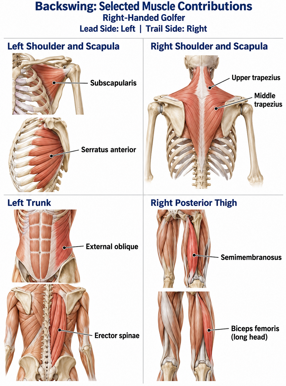
The trail trapezius contributes to scapular positioning during takeaway. EMG research has documented substantial trail-side trapezius activity during this portion of the swing, while the lead serratus anterior remains active across the swing.
Clinically relevant structures include:
Trail trapezius and rhomboids: Contribute to scapular retraction and control as the trail arm moves.
Lead serratus anterior: Helps maintain scapular contact with the thorax while accommodating protraction.
Rotator cuff: Subscapularis, infraspinatus, teres minor, and supraspinatus contribute to glenohumeral control as the arms change position.
Pectoralis major and posterior shoulder tissues: Their loading and extensibility may influence the lead arm’s movement across the chest.
Trunk muscles
The obliques coordinate rotation with the spinal extensors and other trunk muscles. In rightward rotation, the left external oblique and right internal oblique have compatible rotational actions, although actual recruitment also reflects posture, loading, and movement control. Erector spinae and multifidus contribute to controlling spinal position throughout the turn.
Hip and lower-limb muscles
Gluteals, adductors, hamstrings, and quadriceps help support the golfer and control pelvic movement over the legs. Their relevance depends on the position and loading being examined. Trail-side semimembranosus and biceps femoris long head may therefore deserve assessment alongside gluteal function and hip rotation.
Clinical Interpretation
Compare the golfer’s backswing with regional examination:
Restricted turn: Assess active and passive hip rotation, thoracic movement, and how motion is distributed between the pelvis and trunk.
Lead-shoulder symptoms: Examine horizontal adduction, glenohumeral rotation, scapular movement, and relevant resisted loading.
Trail-shoulder symptoms: Assess elevation and external rotation alongside rotator-cuff and scapular-muscle capacity.
Hip, knee, or foot symptoms: Examine the symptomatic region during turning and supported loading, including whether stance or foot positioning changes the response.
Difficulty across repeated swings: Compare initial movement with later repetitions and review practice volume, effort, and recovery.
Separate a movement restriction from difficulty controlling or tolerating the available range. This distinction helps determine the contribution of manual care, exercise, and coaching.
MSR Treatment Priorities and Demonstration
Select relevant myofascial procedures for the scapular muscles, rotator cuff, pectorals, spinal muscles, obliques, gluteals, adductors, or hamstrings when examination connects them with the limitation. Include osseous mobilization or adjustment of relevant joints where indicated.

Phase 1 Demonstration
In the accompanying video, Dr. Abelson demonstrates MSR procedures for selected structures contributing to the backswing. Observe the contact, stabilization, positioning, and movement, adapting procedure selection to the golfer’s findings.
Repeat the golfer’s backswing after treatment using comparable stance, club, and effort. Record changes in symptoms, range, and movement fluidity. Reinforce useful improvements with active rotation, shoulder control, or progressive loading before integrating them into purposeful practice.
The objective is a backswing that prepares the golfer for the next action and remains sustainable across practice and play.

Phase 2: Early Downswing
From the top of the backswing to the club shaft approaching horizontal
Early downswing redirects the body and club toward impact. The golfer must coordinate changing foot pressure, pelvic and trunk rotation, and arm movement while beginning to accelerate the club. For practitioners, this phase helps reveal whether the golfer can use the movement established during the backswing under increasing speed and load.
Anatomy and Biomechanics
In a right-handed golfer, the pelvis turns toward the target as the arms descend. These actions overlap, with their timing and magnitude varying between golfers.
Hips and lower limbs: Support increasingly develops through the lead leg as the pelvis turns leftward. Relative hip rotation shifts toward internal rotation at the lead hip and external rotation at the trail hip. Hip and knee flexion–extension also change as the golfer produces and controls movement.
Pelvis and trunk: Pelvic rotation interacts with thoracic rotation, lateral flexion, and changes in spinal inclination. The trunk must coordinate movement while responding to forces generated through the legs and transmitted through the arms.
Shoulder complex: The arms lower from their elevated backswing position. The lead scapula moves toward retraction, while the trail scapula moves toward protraction. Glenohumeral movement and rotator-cuff activity help control the humeri as the hands redirect the club.
Elbows, forearms, and wrists: The trail elbow begins extending as the arms descend. Forearm rotation and changing wrist positions organize the club’s orientation and developing speed.
The clinical question is whether the golfer can redirect and load the body effectively, including when effort or repetition increases.
Relevant Muscle Contributions
Hip and lower-limb muscles
The trail gluteus maximus and gluteus medius contribute to hip force production and pelvic control. Lead adductor magnus, quadriceps, and hamstrings contribute to supporting and controlling the receiving limb. In competitive golfers, EMG research identified early-downswing contributions from the trail hip extensors and abductors alongside lead adductor magnus, with lead hamstring activity supporting knee control.
Relevant examination targets include:
Trail gluteus maximus and medius: Hip extension, abduction, and control during turning.
Lead adductor magnus: Adductor and hip-extensor capacity in positions relevant to lead-side loading.
Lead quadriceps, including vastus lateralis: Knee-extension capacity and control as loading develops.
Hamstrings: Hip-extension and knee-flexion capacity, interpreted according to the symptomatic side and task.
Trunk muscles
The right external oblique and left internal oblique have rotational actions compatible with turning the trunk toward the target. They coordinate with the remaining abdominal muscles, erector spinae, and multifidus to manage rotation and spinal loading. Their contribution includes controlling movement between the pelvis and thorax.
Shoulder and scapular muscles
Lead middle trapezius and rhomboids contribute to scapular retraction, while trail serratus anterior contributes to protraction as the arms move forward. These changing scapular demands are reflected in golf EMG research.
Pectoralis major and latissimus dorsi contribute to bringing the elevated arms toward the body, according to humeral position. The rotator cuff helps control the glenohumeral joints throughout this transition.

Clinical Interpretation
Connect difficulty during early downswing with regional testing:
Painful lead-side loading: Examine hip rotation, adductor and gluteal capacity, knee movement, and quadriceps loading. Include foot and ankle findings when they affect support or turning.
Difficulty turning from the trail side: Assess trail-hip movement, hip-extensor and abductor capacity, and the relationship between pelvic and thoracic movement.
Shoulder symptoms as the arms descend: Examine relevant shoulder adduction, extension, and rotation, together with scapular movement and resisted loading.
Back symptoms during transition: Compare trunk rotation and lateral flexion with hip movement and loading. Establish whether symptoms change with swing effort or a modified practice task.
Declining control across repetitions: Assess relevant endurance and repeated loading alongside recent practice demands.
A golfer who moves adequately during a slow rehearsal but struggles during a faster transition may require greater loading capacity or coordination rather than additional range.
MSR Treatment Priorities and Demonstration
Select relevant myofascial procedures for the scapular muscles, pectorals, rotator cuff, trunk rotators, gluteals, adductors, quadriceps, or hamstrings when examination connects them with an early-downswing limitation. Coordinate these procedures with osseous mobilization or adjustment of relevant joints where indicated.

Phase 2 - Demonstration
In the accompanying video, Dr. Abelson demonstrates MSR procedures for selected structures contributing to the early downswing. Observe the contact, stabilization, positioning, and movement, adapting procedure selection to the golfer’s findings.
Repeat the transition from the top of the backswing to the club shaft approaching horizontal, using comparable stance, club, and effort. Record changes in symptoms, movement fluidity, and the golfer’s ability to rotate while accepting support through the lead leg. Reinforce useful improvements with appropriate hip loading, trunk control, and purposeful practice.
The objective is a coordinated transition that prepares the golfer to accelerate toward impact and remains repeatable as practice demands increase.
Phase 3: Late Downswing and Acceleration to Impact
From the club shaft approaching horizontal to ball impact
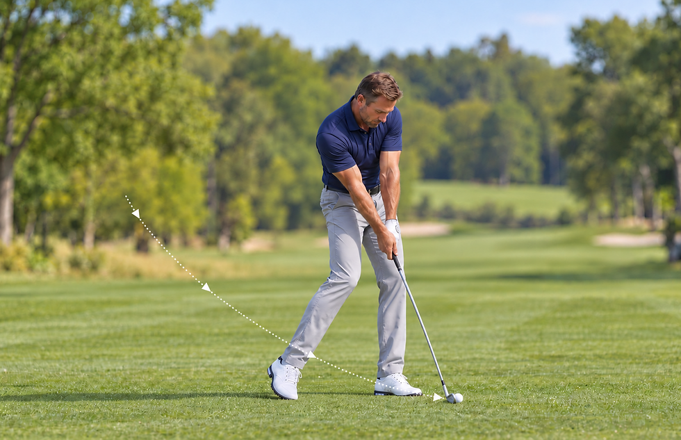
During late downswing, the golfer develops clubhead speed while organizing the clubface for contact. Lower-limb support, pelvic and trunk rotation, shoulder movement, and forearm control must work together within a brief interval. For practitioners, this phase is particularly relevant when greater effort provokes symptoms or the golfer struggles to sustain faster swings.
Anatomy and Biomechanics
In a right-handed golfer, rotation toward the target continues as the hands approach the ball and the clubhead advances through its descending arc.
Hips and lower limbs: The lead leg supports increasing demands as the pelvis turns over it. Lead-knee extension commonly develops approaching impact, while the trail hip and leg continue contributing to movement. Hip rotation, knee control, and foot-ground interaction influence how the golfer manages these forces.
Pelvis and trunk: Pelvic and thoracic rotation interact with changes in spinal extension and lateral flexion. The trunk coordinates force transmission while controlling movement between the pelvis and shoulder girdle.
Shoulder complex: The arms move toward impact through combined glenohumeral and scapulothoracic motion. Lead-arm movement away from the chest accompanies continued trail-arm advancement.
Elbows, forearms, and wrists: Trail-elbow extension and changing forearm and wrist positions contribute to club movement and orientation. Grip requires coordinated finger-flexor activity and wrist-muscle control as external demands increase.
Acceleration results from coordinated segment motion and muscular effort throughout the body. The visible movement of the wrists or arms alone does not explain how the golfer produces speed.
Relevant Muscle Contributions
Pectorals, rotator cuff, and scapular muscles
Pectoralis major and subscapularis demonstrate prominent activity during acceleration in shoulder EMG research. Their contributions should be interpreted alongside humeral position and the movement occurring at each shoulder.
Relevant structures include:
Bilateral pectoralis major: Contributes to humeral adduction and internal rotation, with its mechanical role changing as the arms approach impact.
Subscapularis and the remaining rotator cuff: Contribute to shoulder rotation and glenohumeral control.
Trail serratus anterior: Supports scapular protraction as the trail arm advances.
Lead trapezius and rhomboids: Contribute to scapular retraction and positioning as the lead shoulder moves through the swing.
Research documents differing lead- and trail-side scapular-muscle demands during forward swing and acceleration.
Forearm and hand muscles
Flexor carpi radialis, flexor carpi ulnaris, wrist extensors, pronator teres, and the finger flexors contribute to wrist positioning, forearm control, and maintaining the handle. EMG research has recorded a marked burst of common wrist-flexor activity around contact, highlighting the loading demands in this region.
For examination, distinguish painful gripping from symptoms during resisted wrist movement, forearm rotation, or actual club contact.
Trunk and lower-limb muscles
The obliques coordinate targetward rotation with erector spinae, multifidus, and other trunk muscles. Lead gluteus maximus, quadriceps, hamstrings, and adductors contribute to hip and knee control as the golfer turns over the supporting limb. Trail gluteal and lower-limb contributions continue as support and pressure distribution change.

Clinical Interpretation
Compare the golfer’s acceleration phase with targeted examination:
Symptoms emerging with greater speed: Compare controlled rehearsals with progressively faster swings. Examine relevant force capacity and tolerance to loading at speed.
Lead-hip or knee pain: Assess hip rotation, knee movement, and gluteal, adductor, quadriceps, and hamstring loading in positions relevant to support and turning.
Shoulder or chest symptoms: Examine shoulder rotation, adduction, and horizontal movement alongside pectoral, rotator-cuff, and scapular-muscle capacity.
Elbow, wrist, or hand symptoms: Assess grip, resisted wrist flexion and extension, forearm rotation, and local joint or tendon findings. Distinguish symptoms during acceleration from those triggered by contact with the ball or ground.
Back symptoms approaching impact: Examine relevant trunk rotation, extension, and lateral flexion alongside hip movement and repeated loading.
The timing of symptoms helps refine the clinical question. Pain during an unloaded rehearsal may require a different emphasis from pain occurring only during forceful ball striking.
MSR Treatment Priorities
Consider relevant pectoral, rotator-cuff, scapular, forearm, trunk, and lower-limb findings. Select myofascial procedures and osseous mobilization or adjustment with a specific objective, such as improving shoulder rotation, hip movement, or wrist mobility.
In the accompanying video, Dr. Abelson demonstrates MSR procedures for selected structures contributing to late downswing and acceleration toward impact. Observe the contact, stabilization, positioning, and movement, adapting procedure selection to the golfer’s findings.

Phase 3 - Demonstration
In the accompanying video, Dr. Abelson demonstrates MSR procedures for selected structures contributing to late downswing and acceleration toward impact. Observe the contact, stabilization, positioning, and movement, adapting procedure selection to the golfer’s findings.
Reassess the relevant movement and resisted loading before progressing to faster practice swings or ball striking. Where appropriate, repeat the action from the club shaft approaching horizontal to impact, using comparable club and effort. Connect useful treatment changes with progressive grip and forearm loading, shoulder strength, trunk rotation, and lower-limb preparation.
The objective is to support speed and club control while developing tolerance to repeated acceleration and impact.

Phase 4: Early Follow-Through
From ball impact to the club shaft approaching horizontal
After impact, the body and club continue moving at speed. The golfer must accommodate the remaining momentum while turning over the lead leg and guiding the arms through the next portion of the swing. For practitioners, this phase helps investigate symptoms associated with contact, shoulder rotation, and continued loading through the trunk and lower limbs.
Anatomy and Biomechanics
In a right-handed golfer, the club travels beyond the ball toward the target before rising around the body.
Hips and lower limbs: The lead leg provides substantial support as the pelvis continues turning. Lead-hip rotation and extension develop alongside knee-extension control. The trail heel commonly rises as the trail leg follows the turn.
Pelvis and trunk: Rotation continues while segment velocities change. Muscular activity helps manage this movement and its associated loading; the golfer does not abruptly stop the trunk at impact.
Shoulder complex: The lead arm moves away from its across-chest position as the trail arm advances. Lead-shoulder external rotation and trail-shoulder internal rotation contribute to the developing follow-through, coordinated with scapular movement.
Elbows, forearms, and wrists: The trail elbow continues extending, while forearm rotation and wrist movement accommodate the club’s passage beyond impact. Lead-forearm supination and trail-forearm pronation may accompany this action, with timing influenced by grip and technique.
Shoulder rotation and forearm rotation occur at different joints. Subscapularis and infraspinatus act on the humerus; pronation and supination occur through the radioulnar joints.
Relevant Muscle Contributions
Rotator cuff and pectorals
The trail subscapularis contributes to humeral internal rotation and glenohumeral control, while the lead infraspinatus and teres minor contribute to external rotation and shoulder control. Pectoralis major remains relevant as arm position and loading change. The demand on each structure includes controlling movement as well as producing it.
Shoulder EMG research demonstrates changing recruitment across the swing, including activity associated with follow-through and the extremes of shoulder range.
Scapular muscles
Trail serratus anterior helps accommodate scapular protraction, while lead trapezius and rhomboids contribute to scapular positioning as the chest turns and the arms advance. Research documents sustained trail serratus anterior and lead trapezius activity through the follow-through.
Forearm and hand muscles
Wrist flexors, extensors, finger flexors, and forearm rotators help maintain the handle and manage changing wrist and forearm positions. Contact with the turf or an unexpected obstruction adds a different demand from a practice swing without contact.
Trunk and lower-limb muscles
The obliques, erector spinae, multifidus, and other trunk muscles coordinate continued rotation and changing spinal inclination. Lead gluteals, adductors, quadriceps, and hamstrings contribute to supporting and controlling the golfer over the lead limb.

Clinical Interpretation
Identify whether symptoms begin at contact or develop as the follow-through continues:
Shoulder pain beyond impact: Assess lead-shoulder external rotation, trail-shoulder internal rotation, relevant horizontal movement, and rotator-cuff loading. Compare isolated findings with the arm position that reproduces symptoms.
Elbow or wrist symptoms associated with contact: Examine local joint movement, grip, forearm rotation, and flexor–extensor loading. Ask about turf contact, practice surfaces, and recent changes in striking volume.
Lead-hip or knee symptoms: Assess hip rotation and extension, knee function, and tolerance to supported turning and repeated loading.
Back discomfort during continued rotation: Examine relevant spinal movement alongside lead-hip mobility and trunk capacity.
An abrupt or shortened follow-through: Determine whether pain, restricted movement, fatigue, or a deliberate technical choice explains the pattern.
A slow rehearsal and a full-speed strike provide different information. Select a suitable task that reproduces the concern without repeatedly provoking it.
MSR Treatment Priorities and Demonstration
Consider relevant pectoral, rotator-cuff, scapular, forearm, trunk, and lead-limb findings. Select myofascial procedures and osseous mobilization or adjustment to address the movement or loading limitation identified during examination.
In the accompanying video, Dr. Abelson demonstrates MSR procedures for selected structures contributing to early follow-through, from ball impact to the club shaft approaching horizontal. Observe the contact, stabilization, positioning, and movement, adapting procedure selection to the golfer’s findings.

Phase 4 - Demonstration
In the accompanying video, Dr. Abelson demonstrates MSR procedures for selected structures contributing to early follow-through, from ball impact to the club shaft approaching horizontal. Observe the contact, stabilization, positioning, and movement, adapting procedure selection to the golfer’s findings.
Reassess the passage from impact into early follow-through using comparable stance, club, and effort. Record changes in symptoms, movement fluidity, and tolerance to the relevant loading. Reinforce useful improvements with shoulder-rotation loading, forearm strengthening, trunk control, or supported hip rotation as indicated, then progress toward purposeful ball striking.
The objective is a fluid continuation through impact, with sufficient movement and physical capacity to manage repeated swings.

Phase 5: Late Follow-Through and Finish
From the club shaft approaching horizontal after impact to completion of the swing
The final phase brings the moving body and club toward a controlled finish. Although the ball has already departed, the golfer must still accommodate momentum, complete the turn, and maintain balance over the supporting limb. For practitioners, this phase is useful when shoulder, back, hip, or knee symptoms limit completion of the swing or become more apparent during repeated practice.
Anatomy and Biomechanics
In a right-handed golfer, the pelvis and chest continue turning toward the target as the arms rise and the club travels around the body.
Hips and lower limbs: The lead leg provides the principal support as the pelvis completes its turn. Lead-hip rotation and extension interact with knee position and foot-ground contact. The trail foot commonly pivots onto the forefoot as the heel rises.
Pelvis and trunk: Rotation continues as the golfer becomes more upright. Spinal extension and lateral flexion vary with technique, swing effort, and available hip and thoracic movement.
Shoulder complex: The arms rise and move around the body through coordinated glenohumeral and scapular motion. The lead shoulder moves toward external rotation and horizontal abduction, while the trail arm moves across the chest.
Elbows, forearms, and wrists: Elbow flexion and changing forearm and wrist positions allow the club to wrap around the body as its speed decreases.
A controlled finish does not require every golfer to reproduce the same position. Examine whether the golfer can complete their intended movement without limiting symptoms and sustain that ability across successive swings.
Relevant Muscle Contributions
Rotator cuff, pectorals, and scapular muscles
Lead infraspinatus and teres minor contribute to external rotation and glenohumeral control. Trail subscapularis and pectoralis major contribute according to the position of the trail arm as it crosses the body. Lead pectoralis major also remains relevant to controlling the lead arm’s movement.
Serratus anterior and the trapezius coordinate scapular movement as the arms rise. These relationships make shoulder rotation, elevation, and control useful examination targets when the golfer reports discomfort or difficulty completing the finish.
Trunk muscles
The obliques, erector spinae, multifidus, and other trunk muscles coordinate the remaining turn and the transition toward an upright position. Their contribution includes managing momentum and maintaining balance as movement slows.
Back discomfort in the finish warrants examination of the symptomatic spinal movements alongside hip and shoulder contributions. The visible amount of extension alone does not explain the complaint.
Hip and lower-limb muscles
Lead gluteals, adductor magnus, hamstrings, and quadriceps contribute to support and control as the golfer finishes over the lead limb. Trail gluteal, thigh, and calf muscles accommodate the remaining turn and changing foot contact.
Relevant structures may include lead semimembranosus, vastus lateralis, and adductor magnus, together with bilateral gluteal findings. Their treatment priority depends on the golfer’s examination.

Clinical Interpretation
Connect difficulty completing the swing with regional movement and loading:
Lead-shoulder symptoms: Assess external rotation, horizontal abduction, elevation, and rotator-cuff loading in positions related to the finish.
Trail-shoulder symptoms: Examine across-chest movement, rotation, scapular motion, and relevant pectoral or rotator-cuff loading.
Back discomfort: Assess extension, rotation, and lateral flexion alongside hip mobility and trunk capacity. Compare a controlled finish with the response after a faster swing.
Lead-hip or knee pain: Examine supported turning, hip rotation and extension, knee function, and relevant muscular capacity.
Loss of balance or a finish that deteriorates with repetition: Consider lower-limb support, trunk endurance, fatigue, and swing effort. Coordinate technical questions with the golf professional.
Distinguish reaching the finish position from controlling the movement into it. Comfortable static positioning does not necessarily establish tolerance to the demands of a full-speed swing.
MSR Treatment Priorities and Demonstration
Address relevant rotator-cuff, pectoral, scapular, trunk, hip, or thigh findings through selected myofascial procedures and osseous mobilization or adjustment. Choose treatment according to the movement restriction or loading limitation established during examination.
In the accompanying video, Dr. Abelson demonstrates MSR procedures for selected structures contributing to late follow-through and the finish, from the club shaft approaching horizontal after impact to completion of the swing. Observe the contact, stabilization, positioning, and movement, adapting procedure selection to the golfer’s findings.

Phase 5 - Demonstration
In the accompanying video, Dr. Abelson demonstrates MSR procedures for selected structures contributing to late follow-through and the finish, from the club shaft approaching horizontal after impact to completion of the swing. Observe the contact, stabilization, positioning, and movement, adapting procedure selection to the golfer’s findings.
Reassess the affected finish movement using comparable stance, club, and effort. Record changes in symptoms, movement fluidity, and the ability to complete a balanced finish. Connect useful improvements with shoulder control, trunk rotation, supported hip loading, or balance exercises as indicated. Progress from controlled rehearsals toward the speed and repetition required during practice and play.
The objective is to complete the swing fluidly, manage the remaining momentum, and remain ready for the next shot.
Connecting Phase Findings With MSR Care
The five phases help organize examination, but the golfer performs one continuous swing. A limitation identified during follow-through may involve movement or loading that began earlier. Use the phase to locate the difficulty, then examine the structures and capacities that could explain it.
Establish What Needs to Change
Define the golfer’s priority: greater swing speed, a more fluid turn, improved practice tolerance, or resolution of symptoms during a particular action. Record a reproducible baseline using the relevant club, swing effort, and number of repetitions.
Connect the swing observation with regional testing. Compare active and passive movement, resisted loading, and repeated performance. Palpation can identify tenderness and reproduce familiar symptoms, adding information about where and how to apply care.
Prioritize findings that connect with the golfer’s goal. A measurable restriction matters most when it affects the action the golfer wants to improve.
Combine Myofascial and Joint Care
MSR integrates myofascial procedures with osseous mobilization or adjustment according to the examination. For example:
Restricted backswing: Relevant pectoral or scapular procedures may be combined with shoulder, thoracic, or hip-joint care.
Difficulty loading and turning during downswing: Gluteal, adductor, or thigh findings may warrant treatment alongside relevant hip, knee, foot, or ankle restrictions.
Painful gripping or striking: Forearm procedures may complement wrist or elbow mobilization, with progressive loading directed toward the affected muscle–tendon structures.
Limited follow-through: Rotator-cuff, pectoral, trunk, or hip findings may require coordinated regional treatment.
In my clinical experience, addressing these connected muscle and joint limitations has helped golfers move more fluidly and make better use of their available movement. Application remains specific: contact, stabilization, movement, and intensity should suit the structure, tissue tolerance, and intended change.
Determine Whether the Change Carries Into Golf
Repeat the baseline after treatment. Record whether symptoms, movement, or loading tolerance improve, then introduce an appropriate active task to reinforce the change.
Progress toward the speed and repetition required in practice. Coordinate with the golf professional when the golfer needs help incorporating newly available movement into technique. Subsequent sessions establish whether the improvement remains useful as playing demands increase.
The value of treatment is demonstrated by what the golfer can do with the change: turn more fluidly, develop speed, tolerate repeated strikes, or complete a round with greater confidence.
Conclusion
Understanding the anatomy of the swing helps practitioners explain why a golfer may turn freely during a rehearsal yet struggle with speed, impact, or repeated play. Each phase places changing demands on the shoulders, trunk, hips, and supporting limbs. Connecting those demands with examination makes treatment selection more precise and physical preparation more purposeful.
The MSR approach brings sport-specific biomechanics, myofascial procedures, osseous mobilization or adjustment, and progressive exercise into an individualized plan. The objectives include improving performance, addressing painful limitations, and supporting injury prevention through preparation for the golfer’s intended demands. Progress is judged by the golfer’s ability to use and sustain improvements during practice and play.
In Part 3: MSR Care and Physical Preparation, we develop this application further, connecting hands-on procedures with exercise, practice progression, and ongoing care. The purpose remains practical: helping golfers build the movement, capacity, and confidence to pursue the game they value.
References
Bechler, J. R., Jobe, F. W., Pink, M., Perry, J., & Ruwe, P. A. (1995). Electromyographic analysis of the hip and knee during the golf swing. Clinical Journal of Sport Medicine, 5(3), 162–166. PubMed
Farber, A. J., Smith, J. S., Kvitne, R. S., Mohr, K. J., & Shin, S. S. (2009). Electromyographic analysis of forearm muscles in professional and amateur golfers. The American Journal of Sports Medicine, 37(2), 396–401. pubmed.ncbi.nlm.nih.gov
Glazebrook, M. A., Curwin, S., Islam, M. N., Kozey, J., & Stanish, W. D. (1994). Medial epicondylitis: An electromyographic analysis and an investigation of intervention strategies. The American Journal of Sports Medicine, 22(5), 674–679. pubmed.ncbi.nlm.nih.gov
Kao, J. T., Pink, M., Jobe, F. W., & Perry, J. (1995). Electromyographic analysis of the scapular muscles during a golf swing. The American Journal of Sports Medicine, 23(1), 19–23. PubMed
Lamb, P. F., & Pataky, T. C. (2018). The role of pelvis-thorax coupling in controlling within-golf club swing speed. Journal of Sports Sciences, 36(19), 2164–2171. PubMed
Lephart, S. M., Smoliga, J. M., Myers, J. B., Sell, T. C., & Tsai, Y. S. (2007). An eight-week golf-specific exercise program improves physical characteristics, swing mechanics, and golf performance in recreational golfers. The Journal of Strength and Conditioning Research, 21(3), 860–869. sites.pitt.edu
Marta, S., Silva, L., Castro, M. A., Pezarat-Correia, P., & Cabri, J. (2012). Electromyography variables during the golf swing: A literature review. Journal of Electromyography and Kinesiology, 22(6), 803–813. pubmed.ncbi.nlm.nih.gov
Nesbit, S. M., & Serrano, M. (2005). Work and power analysis of the golf swing. Journal of Sports Science and Medicine, 4(4), 520–533. PubMed
Pink, M., Jobe, F. W., & Perry, J. (1990). Electromyographic analysis of the shoulder during the golf swing. The American Journal of Sports Medicine, 18(2), 137–140. pubmed.ncbi.nlm.nih.gov
Pink, M., Perry, J., & Jobe, F. W. (1993). Electromyographic analysis of the trunk in golfers. The American Journal of Sports Medicine, 21(3), 385–388. PubMed
Vigotsky, A. D., Halperin, I., Lehman, G. J., Trajano, G. S., & Vieira, T. M. (2018). Interpreting signal amplitudes in surface electromyography studies in sport and rehabilitation sciences. Frontiers in Physiology, 8, Article 985. PubMed
Watkins, R. G., Uppal, G. S., Perry, J., Pink, M., & Dinsay, J. M. (1996). Dynamic electromyographic analysis of trunk musculature in professional golfers. The American Journal of Sports Medicine, 24(4), 535–538.
DR. BRIAN ABELSON DC. - The Author

Dr. Brian Abelson is a chiropractor, author, and educator with more than 30 years of clinical experience in musculoskeletal care, rehabilitation, and sports performance. His educational work focuses on musculoskeletal conditions, human movement, sports biomechanics, and evidence-informed approaches to treatment and rehabilitation.
He is the developer of Motion Specific Release (MSR) and Clinical Director of Kinetic Health in Calgary, Alberta, Canada.
Continue Learning with MSR Courses and MSR Pro
For Healthcare Practitioners
Motion Specific Release integrates assessment, clinical reasoning, manual therapy, exercise, and patient education within one adaptable approach to musculoskeletal care.
MSR Pro and MSR courses support different parts of that learning process:
Structured Clinical Education: MSR courses connect applied anatomy and biomechanics with orthopedic and neurological assessment, myofascial procedures, fascial expansions, joint mobilization, manipulation, and rehabilitation.
Hands-On Skill Development: Live training provides opportunities to refine palpation, patient positioning, force application, and clinical decision-making through practice and direct feedback.
MSR Pro Clinical Library: Members receive access to more than 200 MSR procedures, downloadable and fillable clinical forms, examination resources, rehabilitation progressions, and exercise-prescription materials.
Extensive Video Resources: More than 800 videos support examination, treatment procedures, exercise rehabilitation, and clinical application.
Patient Education: A library of 50 condition articles helps practitioners explain common musculoskeletal problems and provide patients with practical information.
Continued Development: Resources are updated as MSR courses and clinical education continue to evolve.
Videos and articles can introduce concepts, but hands-on proficiency develops through deliberate practice, feedback, and experience. MSR is designed to help practitioners connect technique with clinical reasoning and adapt care to the individual rather than apply the same protocol to everyone.
Explore MSR Pro | View Online and Live Courses
Disclaimer:
The articles and embedded videos on the MSR website are provided for educational and informational purposes only. They are not a substitute for individualized assessment, diagnosis, or treatment by an appropriately qualified healthcare professional.
Some videos demonstrate clinical examination and manual therapy procedures intended for licensed or regulated healthcare professionals with appropriate training. These procedures should be performed only within the practitioner’s legal scope of practice and should not be attempted as self-treatment.
Viewing or using this content does not establish a practitioner-patient relationship. If you have questions about a medical condition or symptoms that are new, severe, or worsening, consult an appropriately qualified healthcare provider.
MSR is not responsible for decisions made or actions taken solely on the basis of this educational content. For complete information about website use, third-party links, privacy, and legal terms, please review the MSR Terms of Use, Privacy Policy, and full website disclaimer.


Comments