
Lumbar Spinal Stenosis: Understanding Your Options

Lumbar spinal stenosis (LSS) becomes more common with age. It occurs when the spaces surrounding the nerve roots in the lower spine become narrowed. However, narrowing seen on an X-ray, CT scan, or MRI does not always produce symptoms.
When the nerves are affected, standing or walking may bring on pain, heaviness, cramping, numbness, or weakness in the buttocks and legs. Sitting or leaning forward often provides relief.
The Good News
A diagnosis of lumbar spinal stenosis does not automatically mean surgery. Many people can manage their symptoms and improve their walking capacity through individualized exercise, activity modification, education, and manual therapy when appropriate.
Surgery may be considered when significant limitations persist despite conservative care or when neurological function deteriorates. Because every presentation is different, careful assessment and regular reassessment help determine the most appropriate path forward.
Article Index

Symptoms of Lumbar Spinal Stenosis (LSS)
The hallmark of symptomatic lumbar spinal stenosis is not simply back pain. It is the way symptoms change with standing, walking, sitting, and spinal position.
Common symptoms include:
Buttock or Leg Symptoms: Pain, aching, heaviness, tingling, numbness, or cramping may affect one or both legs.
Reduced Walking Tolerance: Symptoms may gradually increase while walking, limiting how far or how long a person can continue.
Difficulty Standing: Prolonged standing, particularly with the back extended, may intensify leg or back symptoms.
Leg Weakness or Fatigue: The legs may feel tired, heavy, or less dependable during standing and walking.
Balance Changes: Pain, altered sensation, weakness, and reduced confidence may contribute to unsteadiness.
Low Back Pain: Back pain may be present, but some people primarily experience symptoms in the buttocks or legs.
This pattern is known as neurogenic claudication, meaning that standing or walking provokes nerve-related symptoms that improve with sitting or changing position.
Seek Immediate Medical Attention
New loss of bladder or bowel control, numbness around the groin or saddle region, or rapidly worsening leg weakness requires immediate medical assessment. These symptoms may indicate cauda equina syndrome or another serious neurological condition.

How the Spinal Canal Becomes Narrowed
Lumbar spinal stenosis rarely results from one structure alone. More often, several age-related changes gradually reduce the space available for the nerve roots in the central spinal canal, lateral recesses, or openings where the nerves exit.
Common contributors include:
Disc Changes: Spinal discs may lose height or bulge with age, reducing the space around nearby nerve roots.
Facet Joint Changes: The small joints behind the spine can enlarge and develop bone spurs, contributing to further narrowing.
Ligament Thickening: The ligamentum flavum along the back of the spinal canal may thicken or buckle inward, taking up space around the nerves.
Spondylolisthesis: One vertebra may shift relative to another, narrowing the canal or nerve openings.
Developmental Factors: Some people are born with a smaller spinal canal and may develop symptoms with fewer age-related changes.
Spinal position can also influence the available space. Extension, such as prolonged standing, generally narrows the lumbar canal, while sitting or bending forward may increase space and reduce symptoms.
These findings help explain the condition, but anatomy tells only part of the story. Significant narrowing may exist without symptoms, so imaging must always be interpreted alongside the history and clinical examination.

Detective Work: Diagnosing Lumbar Spinal Stenosis (LSS)
Detective Work: Diagnosing Lumbar Spinal Stenosis
Diagnosing symptomatic lumbar spinal stenosis is like assembling a puzzle. No single symptom, examination finding, or scan provides the entire answer. The diagnosis emerges when the history, physical examination, functional pattern, and imaging findings fit together.
Clinical History and Examination
The practitioner will ask what happens during standing and walking, how far you can walk, what relieves the symptoms, and whether you experience pain, heaviness, numbness, or weakness in the legs.
The examination may include strength, sensation, reflexes, gait, balance, hip mobility, and circulation. Because symptoms are often activity-dependent, walking observation or a functional walking test may reveal more than an examination performed entirely at rest.
Imaging and Additional Testing
MRI is commonly used to identify where and how severely the spinal canal or nerve openings are narrowed. CT may be used when MRI is unsuitable or greater bony detail is required. X-rays can show alignment, arthritis, or vertebral slippage but provide limited information about the nerves.
Imaging confirms that narrowing exists, but it cannot determine by itself whether the narrowing is causing the symptoms. EMG or nerve-conduction testing may be considered when the diagnosis remains unclear or peripheral neuropathy is suspected.
Seven Clinical Clues
An international Delphi study identified seven findings that can strengthen clinical suspicion of LSS:
Leg or buttock symptoms while walking
Relief when bending forward
Relief when using a shopping cart or bicycle
Motor or sensory changes while walking
Present and symmetrical foot pulses
Lower-limb weakness
Low back pain
These clues support clinical reasoning, but they do not replace a complete assessment or appropriate imaging.

Differential Diagnosis: What Else Can Look Like LSS?
Several conditions can produce leg pain, numbness, weakness, or difficulty walking. Some may coexist with lumbar spinal stenosis, making careful examination particularly important.
Hip Osteoarthritis: Hip arthritis commonly produces groin, buttock, or thigh pain with weight-bearing. Restricted hip movement and pain during hip testing can help identify its contribution.
Lumbar Disc Herniation: A disc herniation may irritate a nerve root, producing back pain, leg pain, tingling, numbness, or weakness. The symptom pattern may resemble LSS, although it is not always linked to standing and walking.
Greater Trochanteric Pain Syndrome: Pain is typically felt over the outside of the hip and may worsen with side-lying, walking, or climbing stairs. Local tenderness helps distinguish it from spinal pain.
Peripheral Neuropathy: Nerve damage can cause burning, tingling,, numbness, reduced sensation, or weakness, often affecting both feet in a more symmetrical pattern.
Vascular Claudication: Reduced blood flow can cause leg pain or cramping during activity. Symptoms generally improve when activity stops, while neurogenic claudication is more often relieved by sitting or bending forward. Foot pulses and ankle-brachial index testing may assist differentiation.
Cervical or Thoracic Myelopathy: Spinal cord compression higher in the spine can affect gait, balance, coordination, and leg function and requires appropriate medical investigation.
No single feature provides the diagnosis. The overall pattern of symptoms, examination findings, functional testing, circulation, and imaging guides the clinical decision.
Physical Examination: Looking Beyond the Scan
Imaging can show spinal narrowing, but examination reveals how the person moves and whether the clinical pattern matches lumbar spinal stenosis. The practitioner may assess standing tolerance, gait, walking distance, spinal and hip movement, strength, sensation, reflexes, balance, and circulation.

Low Back Examination
A clinical overview of orthopedic and functional procedures used to assess the lumbar spine and identify factors that may be contributing to low back and leg symptoms. No single orthopedic test confirms LSS.

Hip Examination - Orthopaedic Testing Demonstrates inspection, palpation, and active and passive hip movement. These findings help practitioners determine whether the hip may be contributing to symptoms that resemble or coexist with LSS.

Lower Limb Neuro Examination
Reviews muscle strength, sensation, reflexes, and other neurological findings that may indicate nerve-root involvement or another neurological condition.

Peripheral Vascular Examination
Demonstrates key circulation tests used when peripheral arterial disease or vascular claudication is suspected. Abnormal findings may indicate the need for further medical investigation.
These videos are intended for professional education and are not a substitute for an individualized clinical assessment.

Finding the Right Treatment Path for Lumbar Spinal Stenosis
Lumbar spinal stenosis is a common reason for spine surgery in older adults, but a diagnosis does not automatically mean that surgery is required. For many people, the first step is a coordinated nonsurgical program focused on improving walking ability, reducing symptoms, and maintaining independence.
Evidence supports a multimodal approach that may combine education, individualized exercise, activity modification, and manual therapy. These interventions do not reverse the anatomical narrowing, but they can improve mobility, strength, physical capacity, and the way the body manages daily demands.
Manual therapy may include:
Spinal Mobilization or Manipulation: Selected techniques used to address painful or restricted movement in the lumbar spine and related regions.
Soft-Tissue Therapy: Treatment directed toward muscular and fascial tension that may be contributing to pain or limiting movement.
Assisted Mobility: Guided movement for the lumbar spine, hips, and lower limbs based on the patient’s presentation.
Nerve Mobilization: Carefully selected procedures used when neural sensitivity or restricted nerve movement may be contributing to symptoms.
Manual therapy is most useful when it supports active rehabilitation rather than replacing it. Walking or cycling progressions, lower-body and trunk strengthening, balance work, and functional exercise remain central to improving long-term capacity.
The Role of Medication and Injections
Medication may help some patients manage pain, sleep, or participate more comfortably in rehabilitation, but it does not correct the spinal narrowing. Evidence specific to LSS is limited, recommendations differ among guidelines, and potential benefits must be weighed against side effects, age, other health conditions, and medication interactions.
Epidural steroid injections may provide temporary relief for selected patients, but the overall evidence for neurogenic claudication is limited and inconsistent. They should not be presented as a routine solution.
When Surgery May Be Considered
Surgical consultation may be appropriate when pain or walking limitations remain severe despite a reasonable trial of nonsurgical care, or when neurological function is progressively deteriorating. Urgent assessment is required when symptoms suggest cauda equina syndrome or another serious neurological condition.
The best treatment path is not determined by imaging alone. It emerges from the patient’s symptoms, neurological findings, functional limitations, goals, response to care, and informed preferences.
Manipulation and Mobilization: Supporting Movement
The lumbar spine does not move in isolation. The hips, pelvis, thoracic spine, and lower limbs all influence how a person stands, walks, bends, and transfers load. For this reason, treatment may address the painful lumbar region as well as movement restrictions elsewhere in the kinetic chain.
Spinal manipulation and mobilization can play a valuable role in reducing pain, easing stiffness, and helping patients move more comfortably. These techniques do not reverse the anatomical narrowing associated with LSS. Their purpose is to address painful or restricted movement that may be contributing to the person’s overall presentation.
The technique, position, and amount of force should be selected according to the examination findings, neurological status, symptom irritability, bone health, and individual response.

Joint Mobilization - Lumbar Spine
This practitioner-focused video demonstrates lumbar joint-mobilization procedures that may be incorporated into an individualized treatment plan. It shows how controlled, graded movement can be applied to improve mobility and support a more comfortable return to activity.
These procedures require appropriate professional training and should not be attempted as self-treatment.
Soft-Tissue Therapy: Supporting Movement
Muscle tension and restricted soft-tissue movement can add to the discomfort and mobility limitations experienced by some people with lumbar spinal stenosis. Soft-tissue therapy may be used to reduce guarding, improve movement, and help patients participate more comfortably in walking and exercise.
These techniques do not reverse spinal narrowing. They are selected according to the examination findings and used as one part of a broader treatment plan that includes progressive rehabilitation.

Lumbar and Thoracic Spine Fascial Release
The thoracolumbar fascia connects the lower back with the pelvis, trunk, and surrounding muscles. This practitioner-focused video demonstrates fascial-release techniques that may be used when restrictions or tenderness in this region are contributing to pain or limited movement.

Addressing the Deep Paraspinal Muscles
The deep paraspinal muscles help coordinate and support spinal movement. In people with persistent low back pain, these muscles may show changes in activation, endurance, or size. This video demonstrates soft-tissue techniques used to address sensitivity and guarding in the region. Lasting improvements in muscular capacity require appropriately progressed exercise.

Fascial Expansion: MSR Low Back Pain Protocol
This practitioner-focused video features Dr. Brian Abelson demonstrating how MSR fascial-expansion procedures may be incorporated into the care of a patient with low back pain and lumbar spinal stenosis. The approach considers soft-tissue restrictions and movement relationships across the broader kinetic chain.
These procedures require appropriate professional training and should not be attempted as self-treatment.

Exercise: Building Capacity and Confidence
When walking or standing brings on leg pain, heaviness, or weakness, exercise may seem like the last thing you should do. Yet appropriately selected movement is one of the most important ways to maintain independence and improve function with lumbar spinal stenosis.
Exercise does not enlarge the spinal canal, but it can improve:
Walking and standing tolerance
Hip and spinal mobility
Leg and trunk strength
Balance and movement control
Cardiovascular fitness
Confidence with daily activities
The best program is not simply the hardest one. It is the one that matches your symptoms, abilities, health, and goals, then progresses as your capacity improves.

Finding the Right Aerobic Exercise
Walking, cycling, swimming, and water-based exercise can all improve endurance. Because sitting and leaning forward often reduce LSS symptoms, many people initially tolerate a stationary or recumbent bicycle better than upright walking.
Intensity can be guided by the talk test. At a moderate pace, you should be breathing more deeply while still being able to speak in sentences. Heart-rate monitors can provide additional information, but age-based formulas are only estimates and are not required for an effective program.
Begin with a duration you can tolerate, even if that is only 5 to 10 minutes. Increase the time or resistance gradually rather than trying to reach a predetermined target immediately. Several shorter sessions may be more manageable than one long workout.
People with cardiovascular disease, significant balance problems, or other medical concerns should discuss exercise intensity with an appropriate healthcare provider.

Walking
Walking can provoke symptoms, but it is also one of the most meaningful abilities to preserve. Instead of abandoning it, begin with a manageable distance and track how long or how far you can walk before symptoms require you to stop.
A walker, walking poles, or a slight forward lean may make walking more comfortable for some people. Planned rest periods can also help increase total activity.
Temporary symptoms do not always mean harm, but rapidly increasing weakness, worsening balance, or symptoms that do not settle after activity require reassessment. The objective is gradual exposure, not pushing through significant neurological deterioration.
Daily Exercise Routine
The following video demonstrates five movements commonly used to improve hip and spinal mobility in people with lumbar spinal stenosis. These exercises may help some people move more comfortably, but they should be modified according to individual symptoms and physical abilities.
Click Image to Watch Video

5 Best Exercises For Spinal Stenosis
01:10 Pelvic Tilts
02:08 Single Leg Pull
03:10 Fall Over Rotations
04:30 Cat-Cow
05:38 Wide Leg Child’s Pose
Mobility exercises are only one part of rehabilitation. A complete program may also include progressive leg and trunk strengthening, balance training, aerobic conditioning, and practice with meaningful activities such as walking, climbing stairs, lifting, and getting up from a chair.
Stop and seek medical assessment if exercise causes rapidly worsening weakness, saddle numbness, or changes in bladder or bowel control.
Practical Strategies for Daily Life
Small adjustments can make standing, walking, and sleeping more comfortable while you build strength and endurance. The goal is not to avoid every movement that causes discomfort, but to manage symptoms without becoming unnecessarily inactive.

Respond to Symptoms Without Fearing Movement
Activities that repeatedly produce intense or prolonged symptoms may need to be modified temporarily. This could mean shortening the activity, changing position, taking planned breaks, or dividing a task into smaller sessions.
Mild symptoms that settle soon after activity do not necessarily indicate harm. However, increasing leg weakness, worsening balance, or symptoms that persist well beyond the activity should prompt reassessment.

Posture and Extension Sensitivity
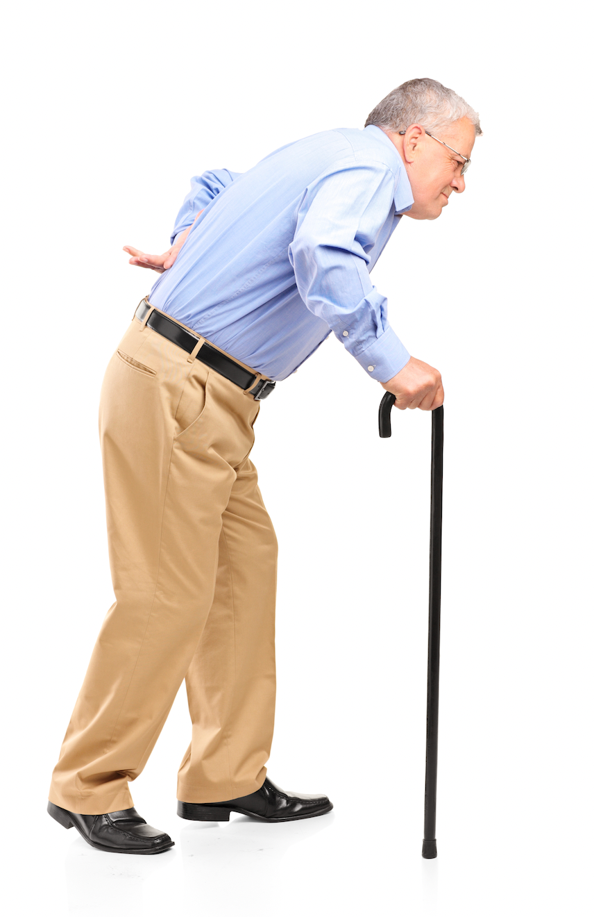
Many people with LSS feel better when sitting or leaning slightly forward because spinal flexion can temporarily increase the space available around the lumbar nerve roots. Standing upright or extending the lower back may narrow that space and provoke symptoms.
Do not force a rigid upright posture if it substantially increases leg pain, numbness, or heaviness. Instead, work gradually toward more comfortable standing and walking through mobility, strengthening, and progressive exposure.
Reaching overhead, walking downhill, or standing for long periods may be difficult for some people, but extension sensitivity is not identical in everyone. Treatment and exercise should be guided by the individual response rather than a universal prohibition against backward bending.
Braces
A lumbar brace may provide temporary comfort or support during selected activities or symptom flare-ups. However, braces do not reverse spinal stenosis, and evidence supporting their routine use is limited.
If a brace is helpful, it should generally complement movement and rehabilitation rather than replace them. Prolonged use should be discussed with a qualified healthcare professional.
Walking Aids
A cane, walking poles, or walker can improve stability and allow some people to walk farther with fewer symptoms. A wheeled walker can be particularly useful because the supported forward-leaning position may reduce neurogenic claudication.
A walking aid is not a sign of failure. It is a tool for maintaining activity, confidence, and independence. Some people need one only during a flare-up, while others benefit from longer-term use. Proper selection and fitting are important.

Finding a Comfortable Sleep Position
No single sleep position works for everyone. Experiment with the following options:
Side-lying with a pillow between the knees
Back-lying with a pillow beneath the knees
Sleeping in a slightly reclined position
Stomach sleeping may aggravate symptoms in people who are sensitive to lumbar extension, but it does not need to be prohibited if it remains comfortable. The best position is the one that allows you to sleep without increasing symptoms.

Our Approach to Lumbar Spinal Stenosis
Lumbar spinal stenosis cannot always be reduced to a single structure or treated with one technique. Symptoms may include anatomical narrowing, nerve sensitivity, restricted movement, reduced strength, changes in balance, and declining walking capacity. Effective care begins by understanding how these factors interact in the individual patient.
Our approach includes:
Comprehensive Assessment: We examine neurological function, walking tolerance, balance, spinal and hip movement, strength, and circulation. Imaging findings are considered alongside the clinical presentation rather than interpreted in isolation.
Individualized Manual Therapy: MSR procedures may be used to address painful or restricted joints and soft tissues throughout the kinetic chain. Manual therapy does not reverse spinal narrowing, but it may reduce discomfort and help patients move and exercise more comfortably.
Progressive Rehabilitation: Exercise programs are designed around the patient’s symptoms, abilities, and goals. Mobility, strength, balance, cardiovascular conditioning, and walking tolerance are progressed as capacity improves.
Practical Self-Management: Patients learn how to modify provocative activities, pace their walking, choose suitable aerobic exercise, and respond constructively to symptom flare-ups.
Regular Reassessment: Progress is measured through meaningful functional changes. When symptoms do not improve, neurological function declines, or the clinical findings raise concern, the treatment plan is reconsidered and medical or surgical referral may be appropriate.
The objective is not to promise a cure or eliminate every symptom. It is to help each person move more confidently, preserve independence, and build as much functional capacity as their condition allows.
References:
Ammendolia, C., Hofkirchner, C., Plener, J., et al. (2022). Non-operative treatment for lumbar spinal stenosis with neurogenic claudication: An updated systematic review. BMJ Open, 12(1), e057724.
Anderson, D. B., de Luca, K., Jensen, R. K., Eyles, J. P., Van Gelder, J. M., Friedly, J. L., Maher, C. G., & Ferreira, M. L. (2021). A critical appraisal of clinical practice guidelines for the treatment of lumbar spinal stenosis. The Spine Journal, 21(3), 455–464.
Bussières, A., Cancelliere, C., Ammendolia, C., et al. (2021). Non-surgical interventions for lumbar spinal stenosis leading to neurogenic claudication: A clinical practice guideline. The Journal of Pain, 22(9), 1015–1039.
Delitto, A., Piva, S. R., Moore, C. G., Fritz, J. M., Wisniewski, S. R., Josbeno, D. A., Fye, M., & Welch, W. C. (2015). Surgery versus nonsurgical treatment of lumbar spinal stenosis: A randomized trial. Annals of Internal Medicine, 162(7), 465–473.
Jensen, R. K., Harhangi, B. S., Huygen, F., & Koes, B. (2021). Lumbar spinal stenosis. BMJ, 373, n1581.
Kalichman, L., Cole, R., Kim, D. H., Li, L., Suri, P., Guermazi, A., & Hunter, D. J. (2009). Spinal stenosis prevalence and association with symptoms: The Framingham Study. The Spine Journal, 9(7), 545–550.
Katz, J. N., Zimmerman, Z. E., Mass, H., & Makhni, M. C. (2022). Diagnosis and management of lumbar spinal stenosis: A review. JAMA, 327(17), 1688–1699.
Lurie, J., & Tomkins-Lane, C. (2016). Management of lumbar spinal stenosis. BMJ, 352, h6234.
Rousing, R., Jensen, R. K., Fruensgaard, S., Strøm, J., Brøgger, H. A., Degn, J. D. M., & Andersen, M. Ø. (2019). Danish national clinical guidelines for surgical and nonsurgical treatment of patients with lumbar spinal stenosis. European Spine Journal, 28(6), 1386–1396.
Schneider, M. J., Ammendolia, C., Murphy, D. R., et al. (2019). Comparative clinical effectiveness of nonsurgical treatment methods in patients with lumbar spinal stenosis: A randomized clinical trial. JAMA Network Open, 2(1), e186828.
Tomkins-Lane, C., Melloh, M., Lurie, J., et al. (2016). ISSLS Prize Winner: Consensus on the clinical diagnosis of lumbar spinal stenosis: Results of an international Delphi study. Spine, 41(15), 1239–1246.
Tomkins-Lane, C., Melloh, M., Wong, A., et al. (2020). Diagnostic tests in the clinical diagnosis of lumbar spinal stenosis: Consensus and results of an international Delphi study. European Spine Journal, 29(9), 2188–2197.
Young, J. J., Hartvigsen, J., Roos, E. M., Ammendolia, C., Kongsted, A., Skou, S. T., Grønne, D. T., & Jensen, R. K. (2021). Symptoms of lumbar spinal stenosis in people with knee or hip osteoarthritis or low back pain: A cross-sectional study of 10,234 participants in primary care. Osteoarthritis and Cartilage, 29(11), 1515–1520.
Disclaimer:
This article and its embedded videos are provided for educational and informational purposes only. They are not a substitute for an individual exami
The exercises shown may not be appropriate for every person. Stop and seek professional advice if an activity causes severe or worsening pain, increasing numbness or weakness, loss of balance, or other concerning symptoms. New bladder or bowel dysfunction, saddle numbness, or rapidly progressive leg weakness requires urgent medical assessment.
Hands-on examination and treatment procedures are intended for appropriately licensed or regulated healthcare professionals with suitable training and must be performed within their professional scope of practice.
Viewing or using this content does not establish a practitioner-patient relationship. Please consult an appropriately qualified healthcare professional regarding your individual condition.
For additional information, please review the MSR website’s full Terms of Use, Privacy Policy, and Disclaimer.
DR. BRIAN ABELSON, DC. - The Author

Dr. Brian Abelson is a chiropractor, author, and educator with more than 30 years of clinical experience in musculoskeletal care, rehabilitation, and sports performance. His educational work focuses on musculoskeletal conditions, human movement, sports biomechanics, and evidence-informed approaches to treatment and rehabilitation.
He is the developer of Motion Specific Release (MSR) and Clinical Director of Kinetic Health in Calgary, Alberta, Canada.

Continue Learning with MSR Courses and MSR Pro
For Healthcare Practitioners
Motion Specific Release integrates assessment, clinical reasoning, manual therapy, exercise, and patient education within one adaptable approach to musculoskeletal care.
MSR Pro and MSR courses support different parts of that learning process:
Structured Clinical Education: MSR courses connect applied anatomy and biomechanics with orthopedic and neurological assessment, myofascial procedures, fascial expansions, joint mobilization, manipulation, and rehabilitation.
Hands-On Skill Development: Live training provides opportunities to refine palpation, patient positioning, force application, and clinical decision-making through practice and direct feedback.
MSR Pro Clinical Library: Members receive access to more than 200 MSR procedures, downloadable and fillable clinical forms, examination resources, rehabilitation progressions, and exercise-prescription materials.
Extensive Video Resources: More than 800 videos support examination, treatment procedures, exercise rehabilitation, and clinical application.
Patient Education: A library of 50 condition articles helps practitioners explain common musculoskeletal problems and provide patients with practical information.
Continued Development: Resources are updated as MSR courses and clinical education continue to evolve.
Videos and articles can introduce concepts, but hands-on proficiency develops through deliberate practice, feedback, and experience. MSR is designed to help practitioners connect technique with clinical reasoning and adapt care to the individual rather than apply the same protocol to everyone.
Explore MSR Pro | View Online and Live Courses


Comments