
Neck Pain: Causes, Diagnosis, Treatment and Exercise

Neck pain is extremely common and can range from a temporary muscle strain to persistent pain associated with the joints, discs, nerves, or other structures of the cervical region. In some cases, symptoms may also be influenced by injury, physical activity, work demands, stress, sleep, and general health.
Because neck pain has many potential causes, effective management begins with understanding what is contributing to the symptoms. A thorough history and physical examination can help identify musculoskeletal problems, neurological involvement, and signs that may require imaging, further investigation, or medical referral.
For many people with musculoskeletal neck pain, treatment may include a combination of education, appropriate activity, exercise, manual therapy, and self-management strategies. Depending on the individual examination, manual therapy may include joint mobilization or manipulation and soft-tissue techniques, while exercise can progressively restore mobility, strength, endurance, and confidence in movement.
This article explores the causes and symptoms of neck pain, clinical examination, imaging, red flags, differential diagnosis, treatment options, and exercise, providing a practical framework for understanding neck pain and making informed decisions about care.
Article Index:
Causes of Neck Pain
Neck pain can develop for many reasons, and sometimes more than one factor is involved. In many cases, symptoms are musculoskeletal and improve with appropriate management. Less commonly, neck pain may reflect neurological, inflammatory, vascular, infectious, or other medical conditions that require further investigation.
Some of the more common causes and contributing factors include:
Muscle and Soft-Tissue Pain: Prolonged or repetitive activities, unfamiliar physical demands, sustained positions, and sudden movements can contribute to muscle fatigue, soreness, or strain. Computer work and frequent phone use may aggravate symptoms for some people, particularly when the neck remains in one position for long periods. Rather than searching for a single “bad posture,” it is often more useful to consider movement variability, physical capacity, and how long a position is maintained.
Injury: Motor vehicle collisions, falls, sports injuries, and other forms of trauma can affect the muscles, ligaments, joints, discs, and other structures of the cervical region. The severity and pattern of symptoms can vary considerably, making an appropriate examination particularly important following significant trauma.
Age-Related and Degenerative Changes: Osteoarthritis, cervical spondylosis, disc degeneration, and spinal stenosis become more common with age. These findings do not necessarily cause pain, and many people have degenerative changes without symptoms. When symptoms and examination findings correspond, however, these changes may be clinically relevant.
Nerve Irritation or Compression: A cervical disc herniation, narrowing around the nerve root, or other conditions can irritate or compress a cervical nerve. This may produce neck pain along with pain, tingling, numbness, or weakness extending into the shoulder, arm, or hand, as can occur with cervical radiculopathy.
Inflammatory, Infectious, Vascular, and Other Medical Conditions: Less commonly, neck pain can be associated with conditions such as inflammatory arthritis, infection, cancer, or vascular disorders. These possibilities are particularly important when neck pain occurs with neurological changes, fever, unexplained weight loss, severe headache, significant trauma, or other concerning symptoms.
Referred Pain: Pain experienced in the neck does not always originate from the cervical spine. The shoulder, temporomandibular joint (TMJ), and other nearby structures can refer pain into the neck. More rarely, medical conditions involving the heart, lungs, or other organs can also produce pain perceived in the neck or shoulder region.
Stress and Psychosocial Factors: Psychological stress does not mean that neck pain is “all in your head.” Stress can influence muscle activity, sleep, physical activity, pain sensitivity, and how symptoms are experienced. For some people, these factors become particularly relevant when neck pain persists or repeatedly returns.
Identifying which of these factors are relevant to an individual is one of the primary goals of a thorough clinical assessment.

Symptoms of Neck Pain
Neck pain can present in many different ways. The location, intensity, duration, and behaviour of symptoms can provide useful clinical information, but no single symptom usually identifies the exact cause. Symptoms should be interpreted alongside the history and physical examination.
Common presentations include:
Localized Neck Pain: Pain may be sharp, aching, or sore and may occur on one or both sides of the neck. The sensation alone does not reliably identify whether the symptoms originate from muscles, joints, discs, or other structures.
Stiffness or Reduced Movement: Some people experience difficulty turning, bending, or extending the neck. This may occur with muscle guarding, joint irritation, injury, or simply because movement has become uncomfortable.
Pain with Movement or Sustained Positions: Symptoms may increase with particular movements or after prolonged activities such as computer work, driving, reading, or looking down at a phone. Understanding which activities aggravate or relieve symptoms can provide valuable information during the examination.
Radiating Arm Pain: Pain extending from the neck or shoulder into the arm may indicate irritation of a cervical nerve root, particularly when it follows a recognizable neurological pattern.
Tingling, Numbness, or Weakness: Neurological symptoms extending into the arm or hand warrant careful assessment. They may occur with cervical radiculopathy or other neurological conditions, but the location and pattern of symptoms need to be evaluated rather than assumed to originate from the cervical spine.
Headache: Neck pain can occur with several types of headache. Cervicogenic headache is associated with dysfunction of structures in the cervical region, but headaches can have many other causes and should not automatically be attributed to muscle tension or cervical degeneration.
Shoulder and Upper-Back Symptoms: Neck pain may occur alongside discomfort around the shoulder blade, upper trapezius, or upper thoracic region. These areas can interact mechanically and neurologically, making examination of the surrounding regions useful.
Dizziness or Balance Disturbance: Dizziness occurring with neck pain deserves careful assessment because it can have vestibular, neurological, cardiovascular, medication-related, and other causes. It should not automatically be attributed to the cervical spine.
The pattern of symptoms helps guide the clinical examination, but diagnosis depends on bringing the history, examination findings, and, when appropriate, further investigation together.
Physical Examination
A thorough clinical examination helps determine what may be contributing to neck pain, identify neurological involvement, and screen for findings that may require further investigation or medical referral.
The examination begins with the patient's history, including how the symptoms started, where they are located, what aggravates or relieves them, whether symptoms extend into the arms or hands, and whether there has been trauma or other concerning symptoms.
The physical examination can then be tailored to those findings and may include observation, palpation, cervical range of motion, orthopedic testing, neurological assessment, and, when clinically indicated, cranial nerve or vascular screening.
No single test establishes the cause of neck pain. The value comes from interpreting the overall pattern of findings alongside the patient's history.
The following videos demonstrate several components that may be incorporated into a neck pain examination.

Cervical Examination
This video provides a structured overview of the cervical examination, including observation, palpation, active and passive range of motion, and selected orthopedic tests.
These findings can help identify painful or restricted movements and guide further examination and treatment planning.

Cranial Nerve Examination
A cranial nerve examination evaluates specific sensory and motor functions associated with the twelve cranial nerves.
It may be appropriate when symptoms such as dizziness, visual disturbance, facial sensory changes, swallowing difficulties, unusual headaches, or other neurological findings raise the need for a broader neurological assessment.

Upper Limb Neuro Exam
An upper-limb neurological examination assesses motor, sensory, and reflex function associated with the cervical nerve roots and peripheral nerves supplying the upper extremity.
This examination is particularly important when neck pain is accompanied by radiating arm pain, numbness, tingling, weakness, or other neurological symptoms. Findings can help determine whether further assessment for cervical radiculopathy, peripheral nerve involvement, or another neurological condition is appropriate.

Vascular Assessment
Vascular assessment may be indicated when the history or symptoms raise concern about circulation or a vascular disorder.
The purpose is not to assume that neck pain has a vascular cause, but to recognize findings that may require additional medical investigation or urgent referral.
Concussion, Dizziness & Vestibular Assessment
Neck pain following a motor vehicle collision, fall, sports injury, or other trauma may occur alongside headache, dizziness, visual disturbance, balance problems, nausea, difficulty concentrating, or other symptoms associated with concussion.
Because cervical, vestibular, neurological, and concussion-related symptoms can overlap, assessment should be guided by the history and clinical presentation.
Dizziness following an injury, for example, should not automatically be attributed to the neck or to concussion. Depending on the symptoms, further neurological, vestibular, or medical evaluation may be appropriate.
The following examinations demonstrate different components that may be used when concussion, dizziness, or vestibular dysfunction is part of the clinical picture.

VOMS
The Vestibular/Ocular Motor Screening (VOMS) assessment is commonly used as part of a broader concussion evaluation. It examines symptom responses during tasks involving eye movements, visual motion sensitivity, and vestibular-ocular function.
VOMS can help identify vestibular or ocular-motor findings that may be relevant following concussion, but it is a screening tool rather than a stand-alone diagnostic test. Results should be interpreted alongside the history and other components of the concussion examination.

HINTS Exam
The HINTS examination, which includes the Head Impulse Test, assessment of Nystagmus, and Test of Skew, was developed to help distinguish certain peripheral vestibular disorders from central neurological causes in patients presenting with acute vestibular syndrome.
Importantly, HINTS is not a general screening examination for dizziness or concussion. It is intended for an appropriate clinical presentation and requires training and experience to interpret correctly. Findings that raise concern for a central neurological cause require urgent medical evaluation.

Dix HallPike Maneuver - Vertigo
The Dix-Hallpike maneuver is used when the history suggests benign paroxysmal positional vertigo (BPPV), particularly when brief episodes of vertigo are triggered by changes in head position.
The maneuver is used to provoke and observe the characteristic vertigo and eye movements associated with posterior-canal BPPV. A positive finding can help identify BPPV and determine whether an appropriate canalith-repositioning procedure should be considered.

Imaging in Neck Pain
Imaging can provide valuable information when the history and clinical examination suggest that further investigation is needed. However, many episodes of uncomplicated neck pain do not require imaging, particularly when there has been no significant trauma, progressive neurological deficit, or other concerning finding.
The decision to order imaging should be based on the clinical presentation and on whether the results are likely to influence diagnosis or management.
Types of Imaging and Diagnostic Testing
X-rays provide information about bones, alignment, fractures, and degenerative changes. They may be appropriate following certain types of trauma or when a bony abnormality is suspected.
MRI provides more detailed visualization of the discs, spinal cord, nerve roots, and other soft tissues. It may be appropriate when there are progressive neurological findings, suspected spinal cord or nerve-root involvement, persistent symptoms that warrant further investigation, or concern about infection, tumour, or other significant pathology.
CT scanning provides detailed images of bone and can be particularly useful in the evaluation of cervical spine trauma or suspected fractures.
Electromyography (EMG) and nerve-conduction studies are not imaging tests. They assess aspects of nerve and muscle function and may sometimes be used when neurological symptoms require further investigation or when the source of weakness, numbness, or radiating symptoms remains unclear.
When Is Imaging Needed?
Imaging is generally most useful when there is a specific clinical reason for obtaining it. Significant trauma, progressive or severe neurological deficits, signs of spinal cord involvement, suspected infection or malignancy, or other red flags may warrant imaging or further medical investigation.
Imaging may also be considered when symptoms persist despite appropriate management and the results are likely to influence the next stage of care.
Avoiding unnecessary imaging is not simply about reducing radiation exposure or cost.
It also helps prevent common age-related findings from being mistaken for the cause of pain when they may be unrelated to the patient's symptoms.
Interpreting Imaging Results
Imaging findings should always be interpreted alongside the patient's history and physical examination.
Disc degeneration, disc bulges, osteoarthritis, and other structural changes become increasingly common with age and can also be found in people without neck pain. Their presence on an X-ray or MRI does not automatically establish that they are responsible for the symptoms.
Conversely, an important imaging finding can significantly influence management when it corresponds with the clinical presentation. For example, evidence of nerve-root or spinal-cord compression that matches neurological findings may alter treatment, prompt further investigation, or lead to specialist referral.
The goal of imaging is not simply to find abnormalities. It is to determine whether an imaging finding helps explain the clinical picture and changes what should be done next.

Identifying Red Flags in Neck Pain
Most neck pain is musculoskeletal and does not reflect a serious underlying condition. However, certain symptoms, examination findings, and elements of the medical history may indicate the need for additional investigation, medical referral, or urgent assessment.
A red flag does not necessarily mean that serious pathology is present. It means that the finding should be considered in the context of the complete clinical picture.
Important findings include:
Progressive Neurological Changes: Increasing weakness, loss of coordination or dexterity, changes in walking or balance, or significant sensory changes may indicate involvement of the spinal cord or nervous system and warrant prompt evaluation.
Signs of Cervical Myelopathy: Hand clumsiness, difficulty with fine motor tasks, gait disturbance, increasing limb weakness, or other upper motor neuron findings can raise concern about cervical spinal cord involvement.
Significant Trauma: Neck pain following a substantial motor vehicle collision, fall, sports injury, or other trauma may require assessment for fracture, instability, neurological injury, or other significant damage.
Fever or Signs of Infection: Neck pain accompanied by fever, chills, severe illness, immunosuppression, or other signs of infection may warrant further investigation for conditions such as meningitis, discitis, osteomyelitis, or spinal epidural abscess.
History of Cancer: New or unexplained neck pain in someone with a history of cancer deserves careful evaluation, particularly when accompanied by other concerning symptoms.
Unexplained Weight Loss or Systemic Symptoms: Significant unexplained weight loss, persistent fatigue, fever, or other systemic symptoms may warrant investigation for underlying medical conditions.
Severe or Unusual Pain: Sudden, severe, rapidly worsening, or unusually persistent pain deserves closer assessment, particularly when the presentation does not resemble the person's previous musculoskeletal symptoms or is accompanied by neurological or systemic findings.
Persistent Night or Rest Pain: Pain at night is common in many musculoskeletal conditions and is not by itself evidence of serious disease. However, severe or progressive pain that remains relatively unaffected by position or activity and occurs alongside other concerning findings may warrant further investigation.
Severe Headache or Neurological Symptoms: Sudden unusual neck pain or headache accompanied by visual disturbance, dizziness, difficulty speaking or swallowing, facial or limb weakness, loss of coordination, or other neurological symptoms requires urgent medical assessment because vascular or neurological causes need to be considered.
Bowel or Bladder Changes: New bowel or bladder dysfunction occurring with neurological symptoms, gait disturbance, or limb weakness requires urgent evaluation for possible spinal cord involvement.
Age and medical history also influence clinical decision-making. Rather than treating “over 50” as a red flag by itself, age should be considered alongside trauma, osteoporosis risk, cancer history, systemic symptoms, neurological findings, and the overall presentation.
Red flags are most useful when interpreted as part of a clinical pattern rather than as isolated warning signs. When serious pathology is suspected, appropriate referral or further investigation should not be delayed.

Differential Diagnosis
Neck pain can arise from many different conditions, ranging from common musculoskeletal problems to less common neurological, inflammatory, infectious, vascular, or systemic disorders. A differential diagnosis is the process of considering these possibilities and determining which are most consistent with the patient's history and examination.
The goal is not simply to identify where it hurts, but to determine what may be producing the symptoms and whether further investigation or referral is needed.
Important categories to consider include:
Musculoskeletal Conditions: Muscle and soft-tissue injuries, cervical joint pain, cervical spondylosis, osteoarthritis, disc-related disorders, and other mechanical causes of neck pain.
Neurological Conditions: Cervical radiculopathy, cervical myelopathy, peripheral nerve disorders, and, less commonly, neurological diseases such as multiple sclerosis.
Traumatic Injuries: Whiplash-associated disorders, muscle or ligament injuries, cervical fractures, and other injuries following motor vehicle collisions, falls, sports injuries, or significant trauma.
Inflammatory and Rheumatologic Conditions: Rheumatoid arthritis, axial spondyloarthritis, polymyalgia rheumatica, and other systemic inflammatory disorders can sometimes involve the cervical region or present with neck and shoulder symptoms.
Infection: Meningitis, vertebral osteomyelitis, discitis, and spinal epidural abscess are uncommon but important conditions to recognize, particularly when neck pain occurs with fever, systemic illness, neurological changes, or relevant medical risk factors.
Vascular Conditions: Cervical arterial disorders, including carotid or vertebral artery dissection, can occasionally present with neck pain or headache and may be accompanied by neurological symptoms requiring urgent medical assessment.
Neoplastic Conditions: Primary tumours and metastatic disease involving the cervical spine are uncommon but should be considered when the history includes cancer or when neck pain occurs with unexplained weight loss, systemic symptoms, progressive neurological findings, or other concerning features.
Referred Pain: Symptoms perceived in the neck may sometimes originate from the shoulder, temporomandibular joint (TMJ), upper thoracic region, or other nearby structures. Less commonly, cardiac, pulmonary, or other medical conditions may produce referred discomfort involving the neck or shoulder region.
Persistent Widespread Pain: Conditions such as fibromyalgia may include neck and shoulder pain as part of a broader pattern of widespread pain, fatigue, sleep disturbance, and increased sensitivity.
Psychological and Social Factors: Stress, anxiety, depression, fear of movement, work demands, and other psychosocial factors can influence pain intensity, disability, recovery, and how people respond to persistent symptoms. Their presence does not mean that the pain is imaginary or purely psychological.
A differential diagnosis develops from the history, physical examination, and pattern of clinical findings. Imaging, laboratory testing, electrodiagnostic studies, or specialist consultation may be appropriate when the presentation warrants further investigation.
The diagnosis should also be reconsidered when symptoms change, neurological findings develop, expected recovery does not occur, or the clinical picture no longer fits the original assessment.
Good clinical reasoning is not simply about identifying the most likely diagnosis. It is also about recognizing when the presentation does not fit.
Treatment of Neck Pain
Treatment should be guided by the diagnosis, severity of symptoms, examination findings, individual goals, and patient preferences. For many types of musculoskeletal neck pain, conservative care may include education, remaining appropriately active, exercise, and manual therapy when indicated.
Some people may also benefit from medical management, particularly when pain is severe, neurological symptoms are present, or another medical condition needs to be investigated or treated. The goal is not simply to reduce pain, but to restore movement and function while progressively rebuilding the capacity to tolerate everyday activities.
Manual therapy can be one component of this approach. Depending on the examination, treatment may include soft-tissue techniques, joint mobilization or manipulation, and other procedures intended to provide short-term improvements in pain or mobility. These changes can create an opportunity for more comfortable movement and active rehabilitation.
Targeting Soft Tissue Injuries

4 Point Neck Pain Protocol
Neck pain does not present exactly the same way in every person. This video demonstrates an MSR approach to assessing and treating soft tissues that may be relevant to an individual's symptoms.
Rather than assuming that one muscle or structure is responsible, treatment should be based on the history and examination and considered within the broader function of the cervical spine, shoulder girdle, thoracic region, and surrounding tissues.
Manual therapy is best used as one component of a broader rehabilitation strategy, particularly when it helps reduce symptoms or improve movement sufficiently to support exercise and normal activity.
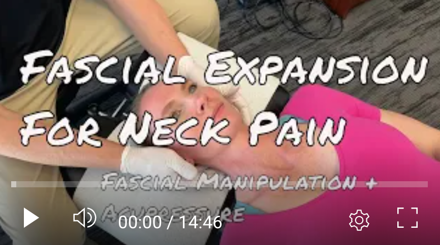
Fascial Expansion: MSR Neck Pain Protocol
The MSR Fascial Expansion approach integrates regional anatomy and biomechanical relationships with selected concepts and points drawn from acupuncture and Traditional Chinese Medicine.
In this video, Dr. Brian Abelson, developer of Motion Specific Release (MSR), demonstrates how Fascial Expansions may be incorporated into the management of neck pain.
This approach should be considered an adjunct to examination, appropriate manual therapy, exercise, and progressive rehabilitation, rather than a stand-alone treatment for all forms of neck pain.
Cervical Mobilization and Manipulation
Joint mobilization or manipulation may be considered when the clinical examination identifies painful or restricted cervical movement and when the procedure is appropriate for the individual.
The goal is not to “put the vertebrae back into place” or permanently correct alignment. These techniques may provide short-term improvements in pain and movement, which can then be reinforced through active movement and exercise.
Cervical Manipulation vs. Mobilization
Both manipulation and mobilization can be used as components of conservative care for selected patients with neck pain. The choice should consider the clinical presentation, potential benefits and risks, contraindications, practitioner judgment, and importantly, the patient's preferences and informed consent.
Mobilization uses slower, controlled movements and may be preferred by patients who do not want cervical manipulation or when manipulation is not clinically appropriate.
Cervical manipulation also requires appropriate screening and clinical reasoning. Rather than using an arbitrary age cutoff, the decision should consider factors such as bone health, significant trauma, neurological or vascular concerns, inflammatory disease affecting cervical stability, structural abnormalities, and other relevant medical conditions.
When contraindications or significant risk factors are identified, another treatment approach should be selected or further medical evaluation obtained.

Cervical Adjustments
This video demonstrates cervical manipulation procedures that may be used by appropriately trained practitioners as one component of care for selected patients with neck pain.
Research suggests that manipulation or mobilization may provide short-term improvements in pain and function for some patients, particularly when combined with exercise and other active approaches. These techniques should not be viewed as universally necessary or as stand-alone treatments for neck pain.

Cervical Mobilization Procedures
Cervical mobilization provides another manual therapy option for addressing painful or restricted neck movement.
In this video, Dr. Abelson demonstrates several cervical mobilization procedures.
The technique selected should reflect the patient's presentation, treatment goals, comfort, and clinical findings.
The objective is not simply to create more movement during treatment. It is to help the patient return to comfortable, confident movement outside the treatment room.
The Role of Exercise in Neck Pain Rehabilitation
Exercise is an important part of rehabilitation for many people with neck pain. Depending on the individual, it can help improve mobility, strength, endurance, physical capacity, and confidence in movement.
There is no single exercise program that is appropriate for every type of neck pain. Exercise should reflect the person's symptoms, examination findings, current physical capacity, and goals. Some people may initially benefit from comfortable mobility exercises, while others can begin strengthening and more demanding activities relatively early.
Manual therapy may be combined with exercise when appropriate, particularly when short-term improvements in pain or mobility make active rehabilitation easier. Over time, however, the goal should be to help the individual become increasingly active and less dependent on passive treatment.
A progressive program may include cervical and thoracic mobility, neck and shoulder strengthening, endurance training, and functional exercises relevant to work, recreation, or sport. General physical activity and cardiovascular exercise can also be incorporated as tolerance improves.
Progression does not need to be completely pain-free. Mild, temporary discomfort during or after exercise may be acceptable for some people, provided symptoms remain manageable and return toward baseline. Exercise should be modified when it produces substantial or progressively worsening symptoms.
The following videos demonstrate several ways that mobility, stretching, and strengthening can be incorporated into a neck rehabilitation program.
Exercise Demonstrations:

5 Minute Neck Pain Relief
This short routine uses gentle movement to help reduce stiffness and encourage comfortable cervical mobility.
It can be used as a starting point for some people with uncomplicated neck discomfort, but the frequency and intensity should be adjusted to the individual rather than automatically performed three or four times each day.

PNF Neck Stretches - Mobility, Strength and Power
Proprioceptive neuromuscular facilitation (PNF) techniques combine stretching with controlled muscular contraction and can be used to develop mobility and neuromuscular control.
In this video, PNF techniques are applied to the cervical region. These exercises may be appropriate for some individuals but should be modified or avoided when they significantly aggravate symptoms or when the clinical presentation requires further assessment.

Neck Strengthening Exercises
Progressive neck strengthening can help improve muscular strength, endurance, and tolerance for physical demands.
Rather than attempting to create a “perfect” posture or guarantee prevention of future neck pain, strengthening should prepare the cervical and shoulder regions for the demands placed upon them during everyday activities, work, recreation, and sport.
The goal of exercise is not simply to make the neck stronger. It is to help the person move with greater capacity, confidence, and independence.
Moving Forward with Neck Pain
Most neck pain can be managed successfully with an appropriate combination of education, activity, exercise, and, when helpful, manual therapy. The key is determining what is relevant to the individual rather than assuming that every case has the same cause or requires the same treatment.
A thorough assessment can help identify neurological involvement, red flags, or other findings that require further investigation, while also guiding rehabilitation for more common musculoskeletal conditions.
The goal is not simply to reduce pain. It is to restore movement, rebuild capacity, and help people return to the activities that matter to them.
References
Blanpied, P. R., Gross, A. R., Elliott, J. M., Devaney, L. L., Clewley, D., Walton, D. M., Sparks, C., & Robertson, E. K. (2017). Neck pain: Revision 2017 clinical practice guidelines linked to the International Classification of Functioning, Disability and Health. Journal of Orthopaedic & Sports Physical Therapy, 47(7), A1–A83.
Kjaer, P., Kongsted, A., Hartvigsen, J., Isenberg-Jørgensen, A., Schiøttz-Christensen, B., Søborg, B., Krog, C., Møller, C. M., Halling, C. M. B., Lauridsen, H. H., Hansen, I. R., Thorsen, H., & Jensen, T. S. (2017). National clinical guidelines for non-surgical treatment of patients with recent onset neck pain or cervical radiculopathy. European Spine Journal, 26(9), 2242–2257.
Chacko, N., Gross, A. R., Miller, J., et al. (2025). Manual therapy with exercise for neck pain. Cochrane Database of Systematic Reviews, 2025(12), CD011225.
Brinjikji, W., Luetmer, P. H., Comstock, B., et al. (2015). Systematic literature review of imaging features of spinal degeneration in asymptomatic populations. American Journal of Neuroradiology, 36(4), 811–816.
Eldaya, R. W., Parsons, M. S., Hutchins, T. A., et al. (2025). ACR Appropriateness Criteria® cervical pain or cervical radiculopathy: 2024 update. Journal of the American College of Radiology, 22(5S), S136–S162.
Borrella-Andrés, S., Marqués-García, I., Lucha-López, M. O., et al. (2021). Manual therapy as a management of cervical radiculopathy: A systematic review. BioMed Research International, 2021.
Mucha, A., Collins, M. W., Elbin, R. J., Furman, J. M., Troutman-Enseki, C., DeWolf, R. M., Marchetti, G., & Kontos, A. P. (2014). A brief Vestibular/Ocular Motor Screening (VOMS) assessment to evaluate concussions: Preliminary findings. American Journal of Sports Medicine, 42(10), 2479–2486.
Kattah, J. C., Talkad, A. V., Wang, D. Z., Hsieh, Y. H., & Newman-Toker, D. E. (2009). HINTS to diagnose stroke in the acute vestibular syndrome: Three-step bedside oculomotor examination more sensitive than early MRI diffusion-weighted imaging. Stroke, 40(11), 3504–3510.
Bhattacharyya, N., Gubbels, S. P., Schwartz, S. R., et al. (2017). Clinical practice guideline: Benign paroxysmal positional vertigo (update). Otolaryngology–Head and Neck Surgery, 156(3 Suppl), S1–S47.
Bialosky, J. E., Bishop, M. D., Price, D. D., Robinson, M. E., & George, S. Z. (2009). The mechanisms of manual therapy in the treatment of musculoskeletal pain: A comprehensive model. Manual Therapy, 14(5), 531–538.
Disclaimer:
The articles and embedded videos on the MSR website are provided for educational and informational purposes only. They are not a substitute for individualized assessment, diagnosis, or treatment by an appropriately qualified healthcare professional.
Some videos demonstrate clinical examination and manual therapy procedures intended for licensed or regulated healthcare professionals with appropriate training. These procedures should be performed only within the practitioner’s legal scope of practice and should not be attempted as self-treatment.
Viewing or using this content does not establish a practitioner-patient relationship. If you have questions about a medical condition or symptoms that are new, severe, or worsening, consult an appropriately qualified healthcare provider.
MSR is not responsible for decisions made or actions taken solely on the basis of this educational content. For complete information about website use, third-party links, privacy, and legal terms, please review the MSR Terms of Use, Privacy Policy, and full website disclaimer.
DR. BRIAN ABELSON, DC. - The Author

Dr. Brian Abelson is a chiropractor, author, and educator with more than 30 years of clinical experience in musculoskeletal care, rehabilitation, and sports performance. His educational work focuses on musculoskeletal conditions, human movement, sports biomechanics, and evidence-informed approaches to treatment and rehabilitation.
He is the developer of Motion Specific Release (MSR) and Clinical Director of Kinetic Health in Calgary, Alberta, Canada.

Continue Learning with MSR Courses and MSR Pro
For Healthcare Practitioners
Motion Specific Release integrates assessment, clinical reasoning, manual therapy, exercise, and patient education within one adaptable approach to musculoskeletal care.
MSR Pro and MSR courses support different parts of that learning process:
Structured Clinical Education: MSR courses connect applied anatomy and biomechanics with orthopedic and neurological assessment, myofascial procedures, fascial expansions, joint mobilization, manipulation, and rehabilitation.
Hands-On Skill Development: Live training provides opportunities to refine palpation, patient positioning, force application, and clinical decision-making through practice and direct feedback.
MSR Pro Clinical Library: Members receive access to more than 200 MSR procedures, downloadable and fillable clinical forms, examination resources, rehabilitation progressions, and exercise-prescription materials.
Extensive Video Resources: More than 800 videos support examination, treatment procedures, exercise rehabilitation, and clinical application.
Patient Education: A library of 50 condition articles helps practitioners explain common musculoskeletal problems and provide patients with practical information.
Continued Development: Resources are updated as MSR courses and clinical education continue to evolve.
Videos and articles can introduce concepts, but hands-on proficiency develops through deliberate practice, feedback, and experience. MSR is designed to help practitioners connect technique with clinical reasoning and adapt care to the individual rather than apply the same protocol to everyone.
Explore MSR Pro | View Online and Live Courses


Comments