
Degenerative Disc Disease: Eliminating Pain, Restoring Function
- Dr. Brian Abelson
- Aug 27, 2024
- 14 min read

Imagine this: 85% of people worldwide sometimes experience lower back pain. This common issue profoundly impacts our daily lives and well-being.
At the core of this discomfort often lies our spinal discs. These complex structures can be significant sources of pain, especially as many of us inherit some degree of disc degeneration, known as Degenerative Disc Disease (DDD). But it’s not just genetics—our environment also plays a crucial role, influencing and sometimes worsening our condition.
The Good News
We have observed a 90% success rate in reducing or eliminating pain and improving function, based on patient outcomes. Combining manual therapy with targeted exercise, as part of our Motion Specific Release (MSR) methodology, can make a real difference in your life. While it may not reverse the progression of Degenerative Disc Disease, it can significantly alleviate pain and enhance functionality.
Article Index

Understanding Degenerative Disc Disease (DDD)
Degenerative Disc Disease (DDD) typically affects the cervical (neck) and lumbar (lower back) regions of the spine. In a healthy spine, intervertebral discs act as shock absorbers between vertebrae. However, with DDD, these discs lose their ability to cushion effectively, leading to significant changes in spinal function.
As the discs degenerate, the spine can suffer from increased wear on the facet joints, often resulting in osteoarthritis. This process can cause spinal discomfort, reduced flexibility, and impaired movement. The body may respond by forming bone spurs (osteophytes) to handle the extra stress, which can further impact spinal health. Over time, the vertebral bodies, especially the end plates, may deteriorate, increasing pressure on the spinal nerve roots and exacerbating pain and dysfunction.

The Vital Role of Intervertebral Discs
Intervertebral discs are essential to our everyday movement and spinal health. These fibro-cartilaginous structures comprise nearly a quarter of the spine’s length and serve as its primary shock absorbers.
Designed to withstand intense biomechanical stress, these discs protect the spinal cord and nerve roots and enhance spinal flexibility, similar to synovial joints.
Each disc is composed of two key parts:
Annulus Fibrosus: The tough outer layer of multi-directional collagen fibres securely attached to the vertebrae.
Nucleus Pulposus: The gel-like core that handles compressive loads, with a high water content crucial for its function.
These discs lack a direct blood supply and rely on nutrient exchange through the vertebral end plates. Disruptions in this process can lead to degenerative changes, affecting overall spinal health. Understanding the role and structure of these discs helps us appreciate their importance in maintaining a healthy, pain-free back.
Decoding Degenerative Disc Disease
It's fascinating to realize that the natural aging of spinal discs is quite different from the changes seen in Degenerative Disc Disease (DDD). Aging and DDD are distinct processes that affect our spines in unique ways.
Let's break it down:
Normal Aging: As we age, the intervertebral discs in our spines naturally lose water content, leading to dehydration by our 40s. However, the discs' height usually remains unchanged. Small fissures may develop as the fibres within the disc become less organized, with aging starting at the disc's center and spreading outward.
Degenerative Disc Disease: Unlike normal aging, DDD causes more specific changes that lead to pain, discomfort, and limited mobility. These differences are crucial for healthcare providers to understand for accurate diagnosis and treatment.

Key Indicators of Degenerative Disc Disease (DDD)
Degenerative Disc Disease (DDD) brings about distinct changes in the intervertebral discs that go beyond normal aging:
Disc Height: A noticeable decrease in disc height weakens its ability to absorb shocks effectively.
Endplate Changes: Unlike normal aging, DDD causes pathological changes in the vertebral endplates, starting from the outer edges and moving inward—a clear sign of the disease.
Rigidity & Weakness: Discs become more rigid and lose strength, reducing their ability to handle stress.
Fissures & Tears: Cracks and tears begin in the outer disc (Annulus Fibrosus) and move inward, allowing nerves and blood vessels to invade, creating pain sources.
Pain Generators: The inward growth of nerves and blood vessels establishes pain generators within the disc.
Fluid Loss: Discs affected by DDD lose fluid faster under pressure, reducing disc height and load-bearing capacity.
In summary, DDD accelerates degeneration, leading to significant structural changes, pain, and reduced function.
Surprising Facts About Cervical Disc Bulges
Cervical disc degeneration is more common than most people think. In a 2015 study by Nakashima and colleagues, 90% of 1,211 healthy volunteers aged 20 to 70 showed disc bulging on MRI scans. Even more surprising, 75% of participants in their 20s had disc bulges. This study highlights that vertebral disc damage can start much earlier than many realize, often beginning in young adulthood.
Diagnosis
In these video demonstrations, you'll see the key orthopedic, neurological, and vascular tests we use to assess patients with suspected Degenerative Disc Disease. These exams are essential for understanding the spine's structure, the health of the nervous system, and any vascular issues that might contribute to symptoms. Performed alongside diagnostic imaging when necessary, these tests help us ensure a thorough and accurate diagnosis.

Orthopedic Testing
This video delves into some typical causes of lower back pain and illustrates how to diagnose them using orthopedic examination techniques. Orthopedic testing is pivotal in the diagnostic process as it helps rule out other potential conditions that might present similar symptoms. By accurately identifying the source of the pain, we can ensure that the patient receives the most suitable treatment for their specific condition.

Lower Limb Neurological Examination
The examination of lower limb neurology plays a significant role in the comprehensive neurological assessment, evaluating the motor and sensory neurons responsible for lower limb function. This assessment is critical for detecting nervous system deficiencies. Neurological testing becomes even more crucial in Degenerative Disc Disease (DDD) due to potential nerve compression, resulting in pain, numbness, or weakness in the lower limbs.

Peripheral Vascular Examination: Key Considerations
A peripheral vascular examination is a crucial diagnostic tool to identify signs of vascular-related conditions. In the case of Degenerative Disc Disease (DDD), this examination takes on added significance. Although DDD primarily affects the spine, it can indirectly affect the vascular system through inflammation or structural changes that may impede blood vessels, potentially causing circulatory problems. Early detection and treatment of Peripheral Vascular Disease (PVD) are vital in preventing cardiovascular and cerebrovascular complications.

Non-Surgical Solutions for Degenerative Disc Disease
Many patients are surprised to learn that Degenerative Disc Disease (DDD) can be effectively managed without surgery. While we can't reverse the degeneration, we can greatly improve their quality of life.
Combining manual therapy with targeted exercises often leads to significant improvements, reducing discomfort and enhancing daily function. Our approach to treating DDD focuses on three key goals:
Enhancing Mobility: Increasing joint movement helps reduce pain and boosts overall functionality.
Reducing Pain and Stiffness: Specialized therapies and exercises alleviate the chronic pain and stiffness associated with DDD, improving your quality of life.
Slowing Disease Progression: While we can't stop the degeneration, our methods can slow it down, helping to maintain joint health longer.
Equally as Effective as Surgery!
Remarkably, ongoing research shows that a combination of manual therapy and exercise can be just as effective as surgery—specifically, spinal fusion—for managing Degenerative Disc Disease (DDD) over time. This non-invasive approach can significantly reduce pain and disability, offering similar outcomes to surgery without the risks and recovery time. It gives patients a powerful alternative, allowing them to choose a treatment path that suits their unique needs and lifestyle while achieving the same level of relief and improved function.
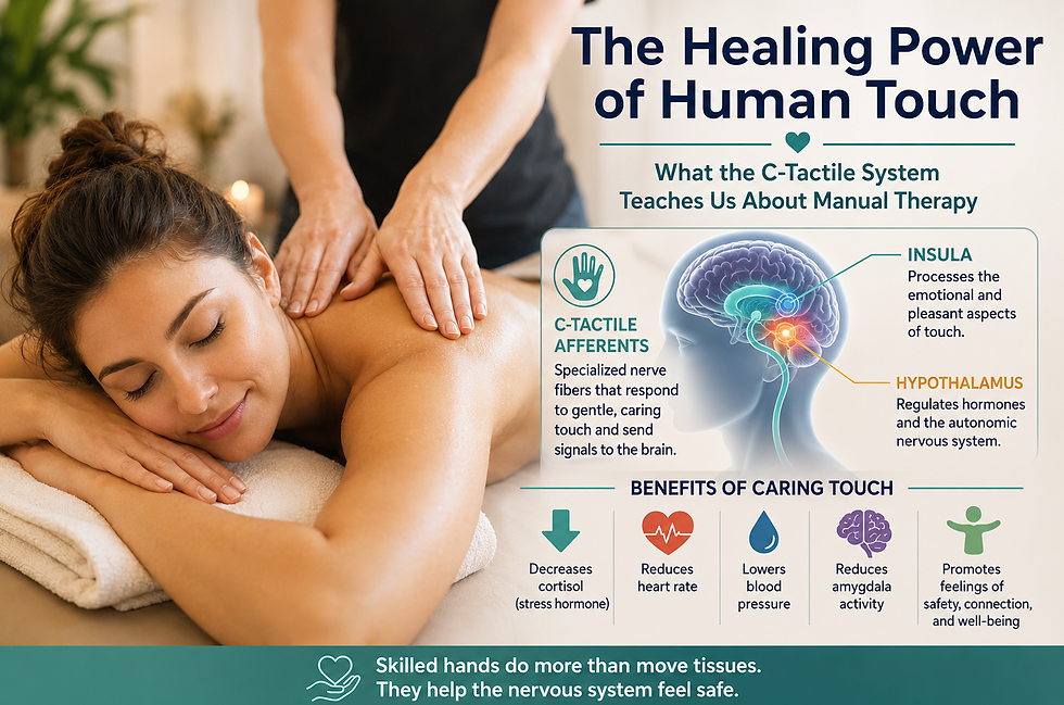
Manual Therapy for Degenerative Disc Disease
Two key factors are essential for achieving the best results when treating degenerative disc disease (DDD) with manual therapy: restoring joint flexibility and improving tissue health.
Joint Flexibility: DDD often leads to stiffness and reduced range of motion in the affected joints. Manual therapy, through hands-on techniques, helps restore lost mobility, ease pain, and enhance overall function.
Tissue Health: DDD can impact the surrounding muscles, ligaments, and tendons. Targeted massage and manipulation in manual therapy improve blood flow, reduce muscle tension, and promote tissue healing, enhancing overall resilience and recovery.

Gentle Chiropractic Care for Degenerative Disc Disease
For patients with chronic back pain from Degenerative Disc Disease (DDD), regular Chiropractic Maintenance Care offers significant relief. Rather than waiting for acute pain episodes, ongoing, scheduled chiropractic treatments can make a real difference.
Through gentle manipulations, even in cases of osteoporosis or severe osteoarthritis, chiropractic care aims to restore spinal function and mobility often compromised by DDD. This approach addresses the affected discs and considers overall spinal health and musculoskeletal balance, helping you move more comfortably and live with less pain.

Cervical Adjustments vs. Mobilization: Your Choice!
When managing Degenerative Disc Disease (DDD) in the neck, you have options: cervical adjustments or neck mobilization.
Cervical Adjustments: These manual techniques restore neck mobility and reduce pain.
Neck Mobilization: A gentler approach that improves flexibility through controlled movements.
Both methods effectively relieve DDD symptoms and enhance neck function, tailored to your comfort and preference. And it's not just the neck—we offer mobilization procedures for the entire spine, ensuring comprehensive care for your spinal health.
Dr. Abelson's video demonstrates Cervical Joint Mobilization and offers insights into the procedure's benefits. Our goal is to provide personalized care that promotes overall spinal well-being.
Soft Tissue Mobilization: Easing the Impact of DDD
In a healthy body, the myofascial system efficiently distributes force and supports smooth motion, acting as a secondary shock absorber for the spine. However, with Degenerative Disc Disease (DDD), this system can become disrupted, leading to muscle imbalances and thickened tissues around the spine, worsening pain and limiting mobility.
Soft tissue mobilization helps counter these effects. Applying targeted pressure and movement to muscles, ligaments, and tendons restores balance, improves tissue health, and enhances spinal stability, relieving DDD-related discomfort.

Low Back Release Protocol (MSR)
n this video, Dr. Abelson demonstrates a low back release protocol designed for general low back pain, which also works effectively for those dealing with Degenerative Disc Disease (DDD).

Fascial Expansion: MSR Low Back Pain Protocol
Fascial expansions offer a powerful approach to managing low back pain, including Degenerative Disc Disease (DDD). This technique integrates modern fascia insights, kinetic chain dynamics, and core acupuncture or traditional Chinese medicine principles. In this video, Dr. Abelson, the Motion Specific Release (MSR) developer, demonstrates how fascial expansions can be used effectively to treat low back pain and relieve those with DDD.
Treatment Frequency for DDD
Effective management of Degenerative Disc Disease (DDD) involves tailoring treatment frequency to the condition's phase:
Acute Phase (Less than 4 Weeks):
Goal: Reduce pain, decrease inflammation, and prevent further damage.
Frequency: Two 15-minute sessions or one 30-minute session per week, with a preference for 30-minute appointments for better results.
Sub-Acute Phase (4 to 12 Weeks):
Goal: Continue healing, restore function, and normalize movement.
Frequency: Manual therapy once a week to support ongoing recovery and address any lingering issues.
Chronic Phase (12 Weeks or Longer):
Goal: Essential maintenance care to reduce or eliminate pain, increase function, and maintain independence.
Frequency: One 30-minute monthly session focusing on maintaining mobility, managing symptoms, and ensuring continued independence.

Exercise: A Key to Managing Degenerative Disc Disease
When managing Degenerative Disc Disease (DDD), certain exercises can help improve mobility, strengthen supporting muscles, reduce pain, and maintain spinal health. Here are some of the best exercises for DDD:
1. Pelvic Tilts
Purpose: Strengthens the lower abdominal muscles and stabilizes the lower back.
How to Do It: Lie on your back with knees bent and feet flat on the floor. Tighten your abdominal muscles and flatten your back against the floor. Hold for a few seconds, then relax.
2. Cat-Cow Stretch
Purpose: Increases flexibility and relieves tension in the spine.
How to Do It: Start on all fours, arch your back towards the ceiling (Cat), then dip your back towards the floor while lifting your head (Cow). Repeat slowly.
3. Bridges
Purpose: Strengthens the glutes and lower back muscles.
How to Do It: Lie on your back with knees bent. Lift your hips towards the ceiling, keeping your shoulders and feet on the floor. Hold for a few seconds, then lower your hips back down.
4. Knee-to-Chest Stretch
Purpose: Stretches the lower back and glutes.
How to Do It: Lie on your back with knees bent. Bring one knee to your chest, holding it with your hands while keeping the other foot flat on the floor. Hold, then switch legs.
5. Lumbar Rotation Stretch
Purpose: Improves spinal mobility and reduces tension.
How to Do It: Lie on your back with knees bent and feet flat. Slowly lower both knees to one side while keeping your shoulders flat on the floor. Hold, then rotate to the other side.
6. Bird-Dog
Purpose: Enhances core stability and strengthens the lower back.
How to Do It: Start on all fours. Extend one arm forward and the opposite leg backward. Hold for a few seconds, then switch sides.
7. Wall Sits
Purpose: Strengthens the legs and stabilizes the lower back.
How to Do It: Stand with your back against a wall, feet shoulder-width apart. Slide down into a seated position as if sitting on a chair, keeping your back against the wall. Hold for as long as comfortable.
8. Standing Hamstring Stretch
Purpose: Stretches the hamstrings and relieves tension in the lower back.
How to Do It: Stand and place one foot on a low stool or step. Keep your back straight, and lean forward slightly until you feel a stretch in the back of your thigh. Hold, then switch legs.
9. Side Planks
Purpose: Strengthens the core, especially the obliques, and stabilizes the spine.
How to Do It: Lie on your side with legs extended. Lift your hips off the floor, balancing on your forearm and the side of your foot. Hold, then switch sides.
10. Low Impact Aerobics
Purpose: Improves overall fitness, increases blood flow, and helps maintain a healthy weight.
How to Do It: Engage in activities like walking, swimming, or using an elliptical machine to avoid high-impact stress on the spine.
These exercises should be performed under the guidance of a healthcare professional, especially if you have DDD, to ensure they are done correctly and safely tailored to your specific condition.
Why Choose Our Approach for Degenerative Disc Disease (DDD) Treatment
While Degenerative Disc Disease (DDD) cannot be fully resolved, our approach consistently achieves a 90% success rate in reducing or eliminating pain and improving function. Here’s why our method is so effective:
Established Expertise: With over 30 years of clinical experience, Dr. Brian Abelson developed the MSR methodology and has successfully treated more than 25,000 patients. You’ll benefit from a proven approach to DDD care.
Thorough Assessments: We conduct detailed evaluations to identify all contributing factors, including nerve compression and muscle imbalances, ensuring a comprehensive understanding of your condition.
Advanced MSR Procedures: Our Motion-Specific Release (MSR) techniques precisely target areas of fascial restrictions, joint dysfunctions, and nerve entrapments, providing practical and lasting relief.
Customized Exercise Programs: We design individualized exercise plans to improve spinal mobility, strengthen supporting muscles, and enhance overall function, helping you regain the ability to live fully and comfortably.
Logical, Evidence-Based Approach: Our treatment protocols integrate manual therapy, exercises, and supportive strategies, ensuring a comprehensive and lasting solution for managing DDD.
Though DDD cannot be cured, our approach helps you manage pain and maintain a higher quality of life. Take the first step toward effective, long-term relief and confidently regain control over your life.
References
Operative and nonoperative treatment approaches for lumbar degenerative disc disease have similar long-term clinical outcomes among patients with positive discography., Smith JS, Sidhu G, Bode K, Gendelberg D, Maltenfort M, Ibrahimi D, Shaffrey CI, Vaccaro AR. World Neurosurg. 2014 Nov;82(5):872-8. doi: 10.1016/j.wneu.2013.09.013. Epub 2013 Sep 15.
Fascia research II. Basic science and implications for conventional and complementary health care., Findley T, and Schleip R. (2009). Introduction. In: Huijing PA, Hollander P, Findley TW, and Schleip R, eds. München: Urban and Fischer.
Fascia: The Tensional Network of the Human Body - E-Book: The science and clinical applications in manual and movement therapy., Schleip R, Findley TW, Leon Chaitow L, and Huijing PA. (2012). Canada: Elsevier
An improved Collagen Scaffold for Skeletal Regeneration, Serafim M. Oliveira, MS, PhD, Rushali A. Ringshia, MS, Racquel Z. LeGeros, PhD, Elizabeth Clark, MS, Michael J. Yost, PhD, Louis Terracio, PhD, and Cristina C. Teixeira, DMD, MS, PhD, J Biomed Mater Res A. 2010 Aug; 94(2): 371–379.
Sakai, D., & Andersson, G. B. (2020). Stem cell therapy for intervertebral disc regeneration: obstacles and solutions. Nature Reviews Rheumatology, 16(4), 213-228.
Nakashima, H., Yukawa, Y., Suda, K., Yamagata, M., Ueta, T., & Kato, F. (2015). Abnormal findings on magnetic resonance images of the cervical spines in 1211 asymptomatic subjects. Spine, 40(6), 392-398.
Thistle, S. (2020). The latest research on musculoskeletal topics. RRS Education.
Maher, C., Underwood, M., & Buchbinder, R. (2017). Non-specific low back pain. The Lancet, 389(10070), 736-747.
Wong, A. Y., Karppinen, J., & Samartzis, D. (2017). Low back pain in older adults: risk factors, management options and future directions. Scoliosis and spinal disorders, 12(1), 14.
Hartvigsen, J., Hancock, M. J., Kongsted, A., Louw, Q., Ferreira, M. L., Genevay, S., ... & Koes, B. W. (2018). What low back pain is and why we need to pay attention. The Lancet, 391(10137), 2356-2367.
Jacobs, W., Van der Gaag, N. A., Tuschel, A., de Kleuver, M., Peul, W., Verbout, A. J., & Oner, F. C. (2013). Total disc replacement for chronic back pain in the presence of disc degeneration. Cochrane Database of Systematic Reviews, (9).
Brinjikji, W., Luetmer, P. H., Comstock, B., Bresnahan, B. W., Chen, L. E., Deyo, R. A., ... & Jarvik, J. G. (2015). Systematic literature review of imaging features of spinal degeneration in asymptomatic populations. American Journal of Neuroradiology, 36(4), 811-816.
Hoy, D., March, L., Brooks, P., Blyth, F., Woolf, A., Bain, C., ... & Buchbinder, R. (2014). The global burden of low back pain: estimates from the Global Burden of Disease 2010 study. Annals of the Rheumatic Diseases, 73(6), 968-974.
Bogduk, N. (2005). Clinical anatomy of the lumbar spine and sacrum. Elsevier Health Sciences.
Ivanov, A. A., Faizan, A., Ebraheim, N. A., & Goel, V. K. (2015). Lumbar degenerative disc disease: current and future concepts of diagnosis and management. Advances in orthopedics, 2012.
De Geer, C. M. (2018). The effect of chiropractic treatment on the reaction and response times of special operation forces military personnel: study protocol for a randomized controlled trial. Trials, 19(1), 425.
Bezci, S. E., Eski, E., & Demir, T. (2020). Effect of manual therapy versus proprioceptive neuromuscular facilitation in patients with chronic degenerative disc disease: a randomized controlled trial. Journal of back and musculoskeletal rehabilitation, 33(1), 143-150.
Kim, S. H., Ahn, S. H., Cho, Y. W., & Lee, D. G. (2013). Effect of intradiscal methylene blue injection for the chronic discogenic low back pain: one year prospective follow-up study. Annals of rehabilitation medicine, 37(5), 675.
Battié, M. C., Videman, T., & Parent, E. (2004). Lumbar disc degeneration: epidemiology and genetic influences. Spine, 29(23), 2679-2690.
Disclaimer:
The content on the MSR website, including articles and embedded videos, serves educational and informational purposes only. It is not a substitute for professional medical advice; only certified MSR practitioners should practice these techniques. By accessing this content, you assume full responsibility for your use of the information, acknowledging that the authors and contributors are not liable for any damages or claims that may arise.
This website does not establish a physician-patient relationship. If you have a medical concern, consult an appropriately licensed healthcare provider. Users under the age of 18 are not permitted to use the site. The MSR website may also feature links to third-party sites; however, we bear no responsibility for the content or practices of these external websites.
By using the MSR website, you agree to indemnify and hold the authors and contributors harmless from any claims, including legal fees, arising from your use of the site or violating these terms. This disclaimer constitutes part of the understanding between you and the website's authors regarding the use of the MSR website. For more information, read the full disclaimer and policies on this website.
DR. BRIAN ABELSON, DC. - The Author

With over 30 years of clinical practice and experience in treating over 25,000 patients, Dr. Abelson created the powerful and effective Motion Specific Release (MSR) Treatment Systems.
As an internationally best-selling author, he aims to educate and share techniques to benefit the broader healthcare community.
A perpetual student himself, Dr. Abelson continually integrates leading-edge techniques into the MSR programs, with a strong emphasis on multidisciplinary care. His work constantly emphasizes patient-centred care and advancing treatment methods. His practice, Kinetic Health, is located in Calgary, Alberta, Canada.

Join Us at Motion Specific Release
Enroll in our courses to master innovative soft-tissue and osseous techniques that seamlessly fit into your current clinical practice, providing your patients with substantial relief from pain and a renewed sense of functionality. Our curriculum masterfully integrates rigorous medical science with creative therapeutic paradigms, comprehensively understanding musculoskeletal diagnosis and treatment protocols.
Join MSR Pro and start tapping into the power of Motion Specific Release. Have access to:
Protocols: Over 250 clinical procedures with detailed video productions.
Examination Procedures: Over 70 orthopedic and neurological assessment videos and downloadable PDF examination forms for use in your clinical practice are coming soon.
Exercises: You can prescribe hundreds of Functional Exercises Videos to your patients through our downloadable prescription pads.
Article Library: Our Article Index Library with over 45+ of the most common MSK conditions we all see in clinical practice. This is a great opportunity to educate your patients on our processes. Each article covers basic condition information, diagnostic procedures, treatment methodologies, timelines, and exercise recommendations. All of this is in an easy-to-prescribe PDF format you can directly send to your patients.
Discounts: MSR Pro yearly memberships entitle you to a significant discount on our online and live courses.
Integrating MSR into your practice can significantly enhance your clinical practice. The benefits we mentioned are only a few reasons for joining our MSR team.


Comments