
The Triceps – An MSR Approach
- Dr. Brian Abelson
- Jan 25, 2024
- 9 min read
Updated: Aug 5, 2024

The triceps brachii muscle is a cornerstone of upper limb anatomy. It serves as the principal extensor of the elbow joint and plays a crucial role in daily functional activities and athletic performance. This article will dissect the intricate anatomy and biomechanics of the triceps brachii within the Motion Specific Release (MSR) framework, emphasizing its central role in arm extension and stabilization.
We will scrutinize the triceps brachii's contribution to elbow extension and its synergistic action with other muscles in the arm, along with the repercussions of its dysfunction on musculoskeletal health. Additionally, we will detail MSR techniques aimed at augmenting the functionality of this muscle group, thereby refining therapeutic interventions for pathologies afflicting the elbow and upper extremity.
Article Index:
In the subsequent sections, we will maintain this structured index to ensure a comprehensive and scientifically detailed exposition of the triceps brachii, suitable for an audience well-versed in medical sciences.

Anatomy & Biomechanics
The triceps brachii, as the primary extensor of the elbow, plays a vital role in the mechanics of the upper limb. A comprehensive understanding of its anatomy and biomechanics is indispensable for implementing effective MSR techniques.
Origin and Insertion:
Long Head Origin: The triceps' long head originates from the scapula's infraglenoid tubercle, crossing both the shoulder and elbow joints.
Lateral Head Origin: The lateral head arises from the posterior surface of the humerus, superior to the radial groove.
Medial Head Origin: The medial head originates from the posterior surface of the humerus, inferior to the radial groove and the entirety of the humeral shaft's medial border.
Insertion: All three heads converge into a single tendon to insert onto the olecranon process of the ulna.
Innervation:
The triceps brachii is innervated by the radial nerve, which is crucial for elbow extension and contributes to the joint's proprioceptive sensation.
Biomechanical Role:
The triceps brachii plays a crucial role in the extension of the elbow joint and is active in various arm positions, contributing to the fine control of arm movements. The long head also assists in adduction and extension of the arm at the shoulder due to its origin on the scapula.
MSR Perspective:
From the perspective of MSR, the triceps brachii is approached as a key element in arm extension and stabilization. MSR techniques focus on improving the triceps' blood flow, elasticity, and strength, while also targeting myofascial restrictions that can hinder muscle function. Understanding the triceps' biomechanical actions enables MSR to aid in the recovery from upper limb injuries, optimize athletic performance, and support the overall functionality of the arm.

Motion Specific Release (MSR) Treatment
Initial Setup:
Patient Position: The patient is seated in a way that provides full access to the tricep muscles.
Practitioner Stance: The practitioner stands in a stable yet dynamic position to apply MSR techniques to the tricep muscles, ensuring full access to the long, lateral, and medial heads.
Basic Technique:
Treatment: The practitioner applies precise hand, or thumb placements and directed pressure along the tricep muscles. The focus is on the long head originating from the infraglenoid tubercle of the scapula, the lateral head above the radial groove, and the medial head below the radial groove on the humerus. Each head can be worked from origin to insertion where needed then reversed.
Support Hand: The opposite hand should stabilize and assist in generating tension with movements, allowing for effective circumduction and traction of the muscle.
Synchronization: The practitioner's movements and pressure should be synchronized to navigate the tricep muscle from its origins to the olecranon process of the ulna, where it inserts.
Pressure Application: Pressure is increased progressively and carefully to ensure patient comfort. Constant feedback from the patient is essential.
Force Generation:
Bilateral Traction: Modulate force in a simultaneous fashion, applying traction both superiorly and inferiorly, to traction the tricep fibers effectively. See demonstration video.
Multidirectional Engagement: Adjust the patient’s arm position to tension the tricep muscle correctly, addressing its complex fiber orientation and fascial connections.
Circumduction Technique: Incorporate circumduction, where the patient assists by moving their arm in a circular motion, while the practitioner applies targeted pressure and follows the movement to enhance the release of fascial restrictions.

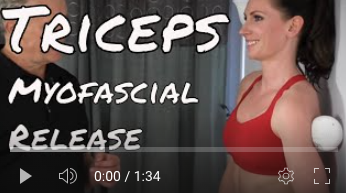
MSR Demonstration Video:
The video includes a demonstration of Dr. Abelson applying MSR to the tricep muscles, showcasing the various techniques, practitioner and patient contact points, and the bilateral traction and circumduction movements that effectively mobilize the muscle.
Best Practices:
Time Allocation: Allocate sufficient time for MSR sessions to ensure a gradual and thorough mobilization of the tricep muscle.
Kinetic Chains: Consider the role of the tricep muscle within the kinetic chain, recognizing its influence on adjacent musculature and overall arm function.
Precautions:
Safety First: Prioritize patient safety, paying close attention to contraindications and obtaining informed consent before beginning treatment.
Gentle Techniques: Apply MSR methods cautiously to avoid exacerbating any pre-existing conditions, maintaining patient comfort throughout the session.
Monitor Patient Feedback: Remain attentive to the patient’s responses, adjusting techniques and pressure accordingly to ensure a responsive and effective MSR session.
Comparative Analysis:
After the treatment, compare the treated arm with the opposite side to evaluate changes in tension and mobility. This comparison provides a clear indication of the MSR treatment's effectiveness.

Functional Kinetic Chains
The triceps brachii's role in upper limb function extends beyond mere elbow extension; its integration within the kinetic chain is vital for coordinated movements and biomechanical efficiency. This integration is mediated through specific myofascial connections and interactions with other musculoskeletal structures.
Direct Myofascial Connections:
Brachial Fascia: This fascial layer envelops the triceps brachii, providing a conduit for force transmission between the upper arm and the forearm, thereby facilitating effective elbow and shoulder movements.
Intermuscular Septa: These fibrous partitions separate the triceps brachii from the surrounding muscles, yet also anchor the muscle to the humerus, aiding in force application during elbow extension.
Synergists:
Anconeus: A small muscle at the elbow that works synergistically with the triceps brachii to extend the elbow joint.
Deltoid: Particularly the posterior fibers, the deltoid assists in shoulder extension and adduction, movements that are complemented by the triceps brachii.
Stabilizers:
Rotator Cuff Muscles: While the triceps brachii are active in shoulder movements, the rotator cuff muscles stabilize the shoulder joint, ensuring a stable platform for triceps function.
Biceps Brachii: The long head of the biceps brachii acts as a stabilizer for the shoulder joint, counterbalancing the forces exerted by the triceps during elbow extension.
Antagonists:
Biceps Brachii and Brachialis: These muscles counter the triceps brachii by providing flexion at the elbow, creating a balanced force dynamic across the joint.
Pronator Teres: While mainly a pronator of the forearm, this muscle also opposes the triceps brachii during elbow extension.
The MSR approach to the triceps brachii acknowledges the complexity of these myofascial interactions and the muscle's role in the functional kinetic chain. By understanding the triceps' connectivity and interplay with surrounding structures, MSR techniques can be finely tuned to address dysfunctions within this network. Such a comprehensive treatment strategy facilitates the restoration of movement efficiency and the integrity of upper limb mechanics.
Exercise
Exercise plays a crucial role in myofascial therapy, aimed at improving flexibility, building strength, and proprioception. Tailored exercises are chosen to match each individual's unique requirements, and the accompanying videos provide examples of potential exercises that may be recommended depending on the case at hand.
Triceps Myofascial Release
Releasing the Triceps muscle can be difficult with the foam roller. In comparison, a softball is ideal to release this muscle.

10 Minute Arm Routine - Dynamic and Isometric
This routine is designed to enhance upper body strength and power by incorporating a combination of dynamic and isometric exercises. These exercises are rooted in anatomy and biomechanics and have been proven to be highly effective.
Conclusion
In the final analysis, the triceps brachii's role extends beyond its basic function of elbow extension to include a broader contribution to the dynamic stability of the upper limb. With this understanding, practitioners can apply focused techniques, including those within the MSR framework, to address specific dysfunctions. Such interventions, informed by a deep understanding of muscle mechanics, aim to restore optimal movement patterns and alleviate discomfort.
The triceps brachii's integration within the kinetic chain highlights the importance of considering the interconnectedness of musculoskeletal components. This perspective is essential in therapeutic practices, where a nuanced approach can lead to significant improvements in function and patient outcomes. The MSR methodology, when applied with an understanding of the underlying anatomy and biomechanics, stands as a valuable tool in the practitioner's repertoire for enhancing musculoskeletal health and mobility.
References
Abelson, B., Abelson, K., & Mylonas, E. (2018, February). A Practitioner's Guide to Motion Specific Release, Functional, Successful, Easy to Implement Techniques for Musculoskeletal Injuries (1st edition). Rowan Tree Books.
Kholinne, E., Zulkarnain, R. F., Sun, Y. C., Lim, S., Chun, J. M., & Jeon, I. H. (2018). The different role of each head of the triceps brachii muscle in elbow extension. Acta Orthopaedica et Traumatologica Turcica, 52(3), 201-205. doi: 10.1016/j.aott.2018.02.005
Kirsch, R. F., Acosta, A. M., Perreault, E. J., & Keith, M. W. (1996). Measurement of isometric elbow and shoulder moments: position-dependent strength of posterior deltoid-to-triceps muscle tendon transfer in tetraplegia. IEEE Transactions on Rehabilitation Engineering, 4(4), 343-352. doi: 10.1109/86.547936
Landin, D., & Thompson, M. (2011). The shoulder extension function of the triceps brachii. Journal of Electromyography and Kinesiology, 21(1), 161-165. doi: 10.1016/j.jelekin.2010.09.005
Maeo, S., Wu, Y., Huang, M., Sakurai, H., Kusagawa, Y., Sugiyama, T., ... & Isaka, T. (2023). Triceps brachii hypertrophy is substantially greater after elbow extension training performed in the overhead versus neutral arm position. European Journal of Sport Science, 23(7), 1240-1250. doi: 10.1080/17461391.2022.2100279
Moore, K. L., Dalley, A. F., & Agur, A. M. R. (2018). Clinically Oriented Anatomy (8th ed.). Philadelphia: Wolters Kluwer.
Netter, F. H. (2018). Atlas of Human Anatomy (7th ed.). Philadelphia: Elsevier.
Santos-Concejero, J., Granados, C., Irazusta, J., Bidaurrazaga-Letona, I., Zabala-Lili, J., Tam, N., & Gil, S. M. (2014). Differences in ground contact time explain the less efficient running economy in north african runners. Biology of Sport, 31(2), 105-112. doi: 10.5604/20831862.1093484
Schoenfeld, B. J., Contreras, B., Willardson, J. M., Fontana, F., & Tiryaki-Sonmez, G. (2014). Muscle activation during low- versus high-load resistance training in well-trained men. European Journal of Applied Physiology, 114(12), 2491-2497. doi: 10.1007/s00421-014-2974-7
Standring, S. (Ed.). (2020). Gray's Anatomy: The Anatomical Basis of Clinical Practice (42nd ed.). Elsevier.
Disclaimer:
The content on the MSR website, including articles and embedded videos, serves educational and informational purposes only. It is not a substitute for professional medical advice; only certified MSR practitioners should practice these techniques. By accessing this content, you assume full responsibility for your use of the information, acknowledging that the authors and contributors are not liable for any damages or claims that may arise.
This website does not establish a physician-patient relationship. If you have a medical concern, consult an appropriately licensed healthcare provider. Users under the age of 18 are not permitted to use the site. The MSR website may also feature links to third-party sites; however, we bear no responsibility for the content or practices of these external websites.
By using the MSR website, you agree to indemnify and hold the authors and contributors harmless from any claims, including legal fees, arising from your use of the site or violating these terms. This disclaimer constitutes part of the understanding between you and the website's authors regarding the use of the MSR website. For more information, read the full disclaimer and policies in this website.
DR. BRIAN ABELSON, DC. - The Author

With over 30 years of clinical practice and experience in treating over 25,000 patients with a success rate of over 85%, Dr. Abelson created the powerful and effective Motion Specific Release (MSR) Treatment Systems.
As an internationally best-selling author, he aims to educate and share techniques to benefit the broader healthcare community.
A perpetual student himself, Dr. Abelson continually integrates leading-edge techniques into the MSR programs, with a strong emphasis on multidisciplinary care. His work constantly emphasizes patient-centred care and advancing treatment methods. His practice, Kinetic Health, is located in Calgary, Alberta, Canada.

Join Us at Motion Specific Release
Enroll in our courses to master innovative soft-tissue and osseous techniques that seamlessly fit into your current clinical practice, providing your patients with substantial relief from pain and a renewed sense of functionality. Our curriculum masterfully integrates rigorous medical science with creative therapeutic paradigms, comprehensively understanding musculoskeletal diagnosis and treatment protocols.
Join MSR Pro and start tapping into the power of Motion Specific Release. Have access to:
Protocols: Over 250 clinical procedures with detailed video productions.
Examination Procedures: Over 70 orthopedic and neurological assessment videos and downloadable PDF examination forms for use in your clinical practice are coming soon.
Exercises: You can prescribe hundreds of Functional Exercises Videos to your patients through our downloadable prescription pads.
Article Library: Our Article Index Library with over 45+ of the most common MSK conditions we all see in clinical practice. This is a great opportunity to educate your patients on our processes. Each article covers basic condition information, diagnostic procedures, treatment methodologies, timelines, and exercise recommendations. All of this is in an easy-to-prescribe PDF format you can directly send to your patients.
Discounts: MSR Pro yearly memberships entitle you to a significant discount on our online and live courses.
Integrating MSR into your practice can significantly enhance your clinical practice. The benefits we mentioned are only a few reasons for joining our MSR team.


Comments